Download
1 / 33
340 likes | 516 Vues
CELLULAR HISTORY & MICROSCOPES. A. Cellular Scientists. Knowledge of cells originated from English scientist Robert Hooke in 1665 Studied thin sections of cork and saw boxlike cavities he called “cells”. Robert Hooke .

E N D
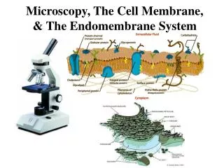
CELLULAR HISTORY & MICROSCOPES
A. Cellular Scientists • Knowledge of cells originated from English scientist Robert Hooke in 1665 • Studied thin sections of cork and saw boxlike cavities he called “cells” Robert Hooke
Dutch scientist Antony van Leeuwenhoek examined pond water in 1676 • observed “animalcules” or tiny animals • Were single-celled amoeba, paramecium, and other water-borne pathogens Water flea daphnia found in pond water Anton van Leeuwenhoek
Matthias Schleiden in 1838 studied plants • cells make up every part of plant – stems, roots, leaves, flowers Matthias Schleiden
Theodor Schwann in 1839 viewed cartilage tissue cells • animals are also made out of cells • Published theory that cells are basic unit of life Theodor Schwann
Rudolf Virchow used work of Schwann and Schleiden to advance cell theory in 1858 Virchow 1821-1902
Cell theory • All living things are made of one or more cells • Cells are basic units of structure and function in organisms • All cells arise from pre-existing cells Apple cells
living organisms may consist of one cell (bacteria) or many cells (plants and animals) that act as a unit or in coordination with each other Staphylococcus aureus Elodea plant
B. MICROSCOPE STRUCTURE • Microscope: instrument used to magnify very small objects 9 1 2 10 3-5 11 6 12 7 13 8 14
C. MAGNIFICATION • Magnification of objective is not total magnification – must consider power of ocular lens, too • Multiply ocular lens by objective lens for correct magnification • Ex: ocular lens has magnification of 4X. If objective lens is 40X, what is total magnification? • 4 x 40 = 160X • Ex: ocular lens has magnification of 10X. If objective lens is 100X, what is total magnification? • 10 x 100 = 1000X
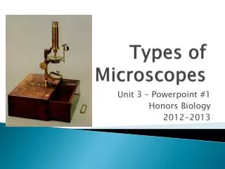
Three types of microscopes, depending on what scientists want to look at: • Light microscope • Electron microscope • Scanning tunneling microscope
Compound Simple
Light Microscope • light microscope: a microscope that uses a beam of visible light passing through one or more lenses to magnify an object up to 1,500 times • Two types: • Simple light microscope (1 lens) • Compound light microscope (2 lenses)
compound light microscope: uses two lenses and a light bulb to shine light up and through object being studied • it contains the following parts: • ocular lens: lens viewer looks through, usually has magnification of 10X • objective lens: lens closest to specimen through which light travels; can have magnification of 20 X, 40X, 100X, up to 200X • specimen: object being studied, usually put on glass slide resting on stage
stage: area specimen rests on, can move up or down to bring object into better focus • focus knob: knob that moves stage up and down to bring object into focus; may have fine focus knob which moves stage at microscopic rate for better resolution • light source: usually a light bulb which shines up through specimen, through objective lens, through ocular lens; may also be a mirror that reflects overhead light
Parts of Compound Microscope MEMORIZE!
to obtain actual magnification, must multiply magnification of ocular lens and objective lens together • if ocular is 10x, and objective is 40x, 10 x 40 = total magnification of 400x 10x 40x (10x)(40x) = 400x
PRACTICE: • If the ocular is 10x and the objective you are using is 10x, what is your total magnification? • 100x • If the ocular is 5x and the objective you are using is 100x, what is your total magnification? • 500x • If the ocular is 2x and the objective you are using is 40x, what is your total magnification? • 80x
Electron Microscope • electron microscope (EM): amicroscope that focuses a beam of electrons to magnify objects up to 200,000 times actual size • two types of EM: • transmission electron microscope (TEM) • scanning electron microscope (SEM)
transmission electron microscope (TEM): electron beam is directed at a very thin slice of specimen stained with metal ions. Heavily stained parts absorb the electrons, while those parts with less stain allow electrons to pass through, and strike a fluorescent screen • these micrographs can reveal cell’s internal structure in fine detail, but are not in color • scientists have to add artificial color to make certain structures more visible
TEM Transmission electron micrograph of myelinated neurons.
SEM This colorized scanning electron micrograph shows pollens—Bermuda grass in green, maple in red, and ragweed in yellow—at roughly 3,000 times their itchy, sneezy life size.
scanning electron microscope (SEM): electron beam is focused on specimen coated with very thin layer of metal. Electrons that bounce off specimen form an image on fluorescent screen. • Shows three-dimensional (3-D) images of cell surface, but are black and white • scientists have to add artificial color to make certain structures more visible
scanning tunneling microscope (STM): uses a needle-like probe to measure differences in voltage caused by electrons that leak, or tunnel, from the surface of the object being viewed • computer tracks movement of probe across object, enabling very small objects the size of an atom to be viewed • computer generates 3-D image of specimen’s surface • can be used for living organisms
STM A scanning microscope (micrograph of the foot of the jumping spider E. arcuata. (Image courtesy Institute Of Physics)
Cell Size • All substances that enter or leave a cell must cross that cell’s surface. • Small cells function more efficiently than larger cells. • About 100 trillion cells in human body • From 5µm-20µm in diameter
Small cells can exchange substances more readily than larger cells because small object have a higher surface area-to volume ratio than larger objects.
Relationship Between Surface Area and Volume Slide length Surface area Volume Surface area/ Volume ratio 1 mm 6:1 2 mm 3:1 4 mm 3:2 If the cell’s surface area-to volume ratio is low, substances can not enter and leave the cell in numbers large enough to meet the cell’s needs.