Download
1 / 46
460 likes | 605 Vues
The Cardiovascular System Chapter 15 http://www.youtube.com/watch?v=yH_Umm37aCc. Cardiovascular System. Heart, blood vessels, blood Arteries, arterioles, capillaries, venules , veins 2 circuits: Pulmonary Circuit Systemic Circuit. The Heart. Pumps 7,000 liters of blood per day

E N D
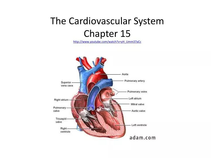
The Cardiovascular SystemChapter 15http://www.youtube.com/watch?v=yH_Umm37aCc
Cardiovascular System • Heart, blood vessels, blood • Arteries, arterioles, capillaries, venules, veins 2 circuits: Pulmonary Circuit Systemic Circuit
The Heart Pumps 7,000 liters of blood per day It takes about 1 minute for blood to circulate around the body Contracts 2.5 billion times in a lifetime Approx. 14cm long and 9 cm. wide
Tissues Covering the Heart • Pericardium – serous membrane that surrounds the heart • Fibrous pericardium (outer layer) • Visceral pericardium (inner layer) • Parietal pericardium (inner lining of fibrous perocardium) • Pericardial cavity – space between the parietal and visceral layers. Contains serous fluid • http://www.youtube.com/watch?v=cnqmfG8rNFE • 54seconds in
Walls of the Heart • Epicardium – visceral pericardium • Myocardium – middle layer, thick and consists mostly of cardiac muscle tissue • Endocardium – inner layer consisting of epithelium and connective tissue
The Mammalian Heart • 4 Chambers – 2 upper atria - 2 lower ventricles • Valves – prevent backflow of blood - tricuspid – R atria, R ventricle - bicuspid – L atria, L ventricle - semilunar – where aorta leaves and the pulmonary artery leaves the heart
Blood Supply to Heart • Coronary Arteries – supply blood to tissues of the heart • The first 2 branches of the aorta • Cardiac Veins – empty into the Right atrium (coronary sinus) http://www.youtube.com/watch?v=gIXcWE0bTwY
Heart Attack • A block or narrowing of a coronary artery or one of its braches deprives myocardial cells of oxygen • Angina Pectoris – may occur during activity or physical stress • A complete blockage kills heart tissue = Myocardial infarction (MI) or a heart attack
The Cardiac Cycle • The heart beats in a coordinated fashion • Atrial Systole – contraction of the atria • Ventricular Diastole – relaxation of the ventricles • Cardiac Cycle – contraction and relaxation of the heart • Pressure in heart rises and falls which opens and closes the heart valves Ex: ventricles contract causing pressure to rise, when it exceeds atrial pressure, AV valves close. Papillary muscles contract, pull on chordae tendineae, and keep tension on valves. Atria are now relaxed, causing them to fill with blood.
Sounds of the Heart • “lubb-dupp” • Lubb = ventricular systole, AV valves closing • Dupp = ventricular diastole, pulmonary and aortic valves closing • http://depts.washington.edu/physdx/heart/demo.html
The Heart Conduction Systemclumps/strands of specialized cardiac muscle tissue • Sinoatrial (SA) node : maintains rhythm - pacemaker - found in wall of R atrium where the sup vena cava enters • Atrioventricular (AV) node - found in wall between R atrium and R ventricle in the interatrial septum - delayed about 0.1 sec, allows atria to fully contract and empty
Cardiac impulses pass from SA Node down junctional fibers to the AV Node • Impulse passes from AV Node to the AV Bundle (atrioventricular bundle or bundle of His) • ½ down the septum – Purkinje fibers take impulses down the heart, spread into papillary muscles and to the apex of the heart. Then pass upward along the lateral walls. They branch off along the way
Intercalated disks – anchors muscle cells together. Gap junctions are found in these disks and allow communication from one cell to another.
ECG • Records the electrical currents of the heart
Arteries and Arterioles • Elastic vessels composed of 3 layers (tunics) 1. tunica interna inner, squamous epithelium 2. tunica media middle, smooth muscle fibers 3. tunica externa (adventitia) outer, connective tissue
Capillaries • Thin walls allow for exchange of materials (semipermeable) • Contain intercellular slits – liver, spleen, red bone marrow have largest slits • Capillaries are densest in muscle and nerve tissue, least dense where metabolism is lowest
Regulation of Capillary Blood Flow • Regulation of blood flow into capillary beds controlled by smooth muscle sphincters which open and close in response to demands placed upon it • Controlled by oxygen levels – low O2, sphincter relaxes, blood flow increases
Exchange in Capillaries • Diffusion of lipid soluble substances – O2, CO2, fatty acids • Lipid insoluble – water, Na+, Cl- - through pores and slits • Hydrostatic pressure pushes materials through membrane
Venules and Veins • Carry blood back to atria • Follow similar paths as arteries • Walls composed of 3 layers, however, thinner walls than arteries with less smooth muscle • Many contain flaplike valves – semilunar valves
Pressure is minimal in veins: How does blood flow through veins? 1. Closed system 2. Activity of skeletal muscles and valves 3. Contraction of smooth muscle in vein walls 4. Pressure changes in thorax when we inhale
Normal blood pressure = 120/80 systolic – pressure during 1 ventricle contraction diastolic – pressure during relaxation of heart 140/90 = hypertensive 90/60 = hypotensive
Cardiac OutputThe volume of blood discharged from the ventricle per minute • Cardiac Output = stroke volume x heart rate stroke volume = volume of blood pumped by ventricle per beat heart rate = # of times the heart beats per minute CO = (70 ml of blood) x (72 bpm) CO = 5,040 ml blood per minute • Blood volume = about 5 liters for adults 8% of body weight
Peripheral Resistance • Resistance between blood and the walls of the blood vessels • Factors Affecting: Contraction of smooth muscles increases resistance Dilation decreases resistance Viscosity – resistance to flow Blood cells and plasma proteins increase viscosity
Control of Blood Pressure • BP = CO x PR (peripheral resistance) • Only about 60% of the end diastolic volume is pumped out of the ventricle in a normal contraction • As blood enters ventricles, myocardial fibers as stretched – PRELOAD - the greater the end-diastolic volume (EDV), the greater the preload = greater the force of contraction Frank-Starling law of the heart – relationship between fiber length and force of contraction
Blood • 4 – 6 L of blood in average human • Plasma – liquid matrix, 90% water pH 7.35 – 7.45 plasma proteins act as buffers, maintain osmotic balance, contribute to viscosity
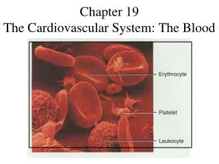
Cellular Components of Blood • RBC’s (erythrocytes) – 25 trillion (5-6 million/mm3) lack nuclei generate ATP anaerobically function to carry oxygen Each cell – 250 million hemoglobin molecules
Production of RBC’s • Negative feedback, sensitive to O2 • Decrease of O2 increase of erythropoietin (done in kidney) which stimulates RBC formation
White Blood Cells (leukocytes) • 5 types 1. Monocytes 2. Neutrophils 3. Basophils Fight infection 4. Eosinophils 5. Lymphocytes • 5000 – 10,000 mm3
Platelets • Fragments of bone marrow cells • Function in blood clotting