Download
1 / 43
470 likes | 982 Vues
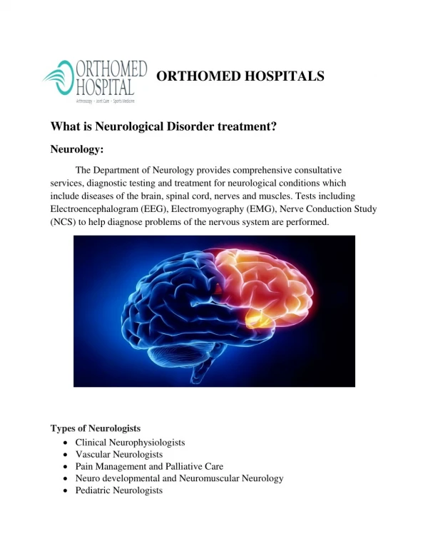
Common Diagnostic Procedures in Neurology. superKAT :) . Neuroimaging. Diagnostic imaging techniques Rapidly growing Increasing important in chronic and acute stages stroke Accuracy is essential . CT Scan – Computed Tomography. Provide “slice” images of brain and spinal cord

E N D
Common Diagnostic Proceduresin Neurology superKAT :)
Neuroimaging Diagnostic imaging techniques • Rapidly growing • Increasing important in chronic and acute stages stroke • Accuracy is essential
CT Scan – Computed Tomography • Provide “slice” images of brain and spinal cord • Axial X-ray beams pass through the head • Amount of radiation is essentially harmless • Degree to which the tissue attenuate the X-ray beams are primarily important for differentiation
Computed tomography • X-ray attenuation of the skull, CSF, cerebral gray and white matter, blood vessels • 30,000 beams of x-ray directed at several axial or coronal levels • Differing densities of bone and intracranial
Indications • Infraction • Hemorrhages, AVM, aneurysm • Edematous tissue • Changes in cranial structures Useful for patients are neurologically/medicaaly unstable, uncooperative, clasutrahoibc with pacemakers
CT angiography • Widely available • Less Specialized skill requirement • Less invasive intravenous administration
Kinds of CT • Contrast enhanced CT (CECT) • Detect lesions that involve breakdown of blood brain barrier • Used to rule out brain tumors and abscesses • Intravenous contrast medium -based on iodine
CT findings • Low attenuation – appear black • Air (darkest) • Fat • CSF and water • Medium attenuation – Gray • Edematous/infraction • Normal brain • Subacute hemorrhage • White • Hemorrhage • Intravenous contrast material • Bone or metal
Appearance of ICH Hypertensive bleed • Occurs disruption of small arteries of thalamus and putamen • Acute or chronic hypertension • Located within the basal ganglia and pons
Appearance of ICH • Hyperdense in all kinds of acute ICH • High hematocrit (90%) in hematoma after clotting and retraction • High density of protein component of Hgb • Mass effect of hematoma maybe present • Extended hypodense ring around an ICH may indicate perifocal edema (rare in the 1st 6 hours)
Subarachnoid Hemorrhage • Spontaneous rupture of aneurysm of the basal cerebral arteries • Hemorrhage into subarachnoid space
Appearance of SAF • Non contrast CT scan detect even small amounts of subarachnoid blood • Overall sensitivity is 92% to 98 within the first 24 hours after SAH • Gold standard is still lumbar puncture (drawing CSF from subarachnoid space)
Appearance of SAF • Location does not necessarily predict origin except in combination with intracerebral hemorrhage • Can be misleading if ICH is large and aneurysm rupture is not considered
Acute ischemic stroke • Result from the thrombo-embolic occlusion of intracranial arteries • Various patterns that can be seen in CT scan in early and late stages of the stroke
Anterior cerebral artery • Isolated infract is rare • Local angiopathies such as vasculitis • More frequent found in carotid T-occlusions (ACA or MCA) • Following multiply cardioembolic events
CT findings • Early • Clear visible hypodense area in cortical rim which may be missed occasionally • Late • Unusual to have difficulty in recognizing hypodensity of a lesion • Increase tissue contrast compared with normal brain tissue
Middle cerebral artery (MCA) • Most common vessel in ischemic strokes = 75% of all • Symptoms vary between minor sensory or motor deficits • More patients scanned earlier to rule out hemorrhage and large infracts due to thrombolytic therapy
Hyperdense MCA signs (HMCAS) • Clot is visible as hyperdense in MCA • Most evident in the horizontal part in the MCA • Appears as a vessel segment of higher density than other parts of same vessel, contralateral MCA and BA • Not an unequivocal sign of occlusion • Does not represent ischemic changes in parenchyma
CT scan • Advantages Disadvantages • Easilly detecting: • Parenchymal bleed posterior circulation vascular disease • SAH, IVH bone related artifacts • Pressure effects limited views • Aneurysm suboptimal brain resolution • Readily available • Less expensive
Complications • For contrast phase : caution among patients with renal problmes • Normal creatinine level
Magnetic resonance imaging • Images displayed as maps of tissue signal intensity values • Spatial localization
Provides thin clice images of the brain and spinal cord • Better resolution • Magnetic field aligns the protons of tissues and CSF in the orientation of the field • Radio frequency pulse causes the proton to resonate and chande their axis
Indications • Hemorrhages • Ischemic/occlusive strokes (lacunes, brainstem and cerebellum) • Demyelinating disease • Tumors • Hypertensive encephalopathy • Vascular anomalies
Advantages Disadvantages • Select any plane Claustraphobia • Does not need radiation bone imaging limited to display of marrow only • No bone artifacts cannot use with pacemakers no ferromagnetic implants • Brainstem lesions
T1 images • Best for showing anatomy • CSF and bone appears black • Normal brain is gray • Flat and subacute hemmorhage appear (>48hours) white
T2 images • Best for showing pathology • CSF and brain appear white • Normal brain=gray • Bone will appear black
MR angiography • Prodcue images of intracranial and extracranial cerebral circulations • Adequate doe evaluation of large lesions • MR venography • Provides subtraction images of major venous sinuses • Useful in dural sinus thrombosis • Less sensitive that angiography
Angiography • Provide high resolution images of the extra intra cranial cerebaral vasculature • Small cathther is threaded into extracranial vessels through the femoral artery
Angiography – 4 vessel • Functional imaging test • Injection of contrast material • Flowing blood to produce signals • Occlusions, stenosis (narrowing), anuerysm • angitis
Identifying the following • Occluded or stenotic vessel • Arterial dissection • Aneurysm • AVM • Vasculitic narrowing • Venous sinus thrombosis
complications • Stroke • Most important complication • 1-2% • Results in emboli generated by catheter • Occurs more frequently in elderly with atherosclerosis dieses 2. Bleeding
Transcranial Doppler (TCD) • Detects blood velocities (pattern of blood flow) • Temporal and Suboccipital window (VA, BA) • Temporal window (post cerebral, PCOM)
ACA • MCA • BIFUR – siphon • PCA • BA • VERTebral artery • OPH • SIPHON – internal carotid hook up give branches to MCA and ACA
sensitivity • Detect for occlusions • Stenosis
EEG – Electroencephalography • Scalp electrodes • Examines spontaneous electrical activity of the brain • Tiny electrical potentials, measured as microvolts (uv), are recorded, amplified and displayed
Preparations • Scalp clean • Precipitating activities • Induced drowsiness or sleep • Stroboscopic retinal stimulation (strobe lights)
Indications • Epilepsy • Suspected seizure disorder • Toxic diseases – lead • Metabolic diseases – hypocalcemia, renal failures • Sleeping disorders – insomnia, sleep study • SSPE – subscleroting (?) pan encephalitis (widespread encephalitis), reactivation of measles virus, 9/10yrs until adolescence
Lumbar puncture • To get CSF sample for diagnosis and treatment purposes, spinal needle – medium sized • Interspace: L4-L5 (infants), adults: L3-4 • Side lying position • Fetal like position • ASIS – anatomic landmark • Ensure bleeding parameters
Preparations • Aseptic technique • Spinal needle • Test tubes • Manometer (pressure readings = opening and closing) • Sterile sheet • Sterile gloves • Cotton and Betadine • Band-aid, gauze
Indications • Pressure measurements • Procure sample of CSF • Cellular, chemical and bacteriologic examination • Administer antibiotics, chemotherapeutic agents, spinal anesthetics • Detecting disease: • CNS infection (Meningitis) • Subarachnoid hemorrhage (SAH) • Neoplasms (no signs of impending herniation)
CSF pressure measurement • CSF collection • CSF appearance – traumatic vs SAH • CSF analysis • Bacteriological (RBC, WBC, gm stain, AFB, india ink) • Biochemical (protein and glucose) • Special tests (Cell cytology – extension of cancer in the CNS, LJ culture – Lowenstein Jensen detect presence of CB, CALAS, oligoclonal bands – demyelinating process , Sabauraud’s culture)
Normal Opening pressure = 80-180mm H20 RBC = 0-5cumm ideally none WBC = 0-5cumm all lymphocytes
NPO and flat on bed for 4 hours • Complications: • Headache • Radicular pain • Vomiting