Download
1 / 42
430 likes | 764 Vues
OBSTRUCTIVE AIRWAYS DISEASE &. SMOKING-ASSOCIATED INTERSTITIAL LUNG DISEASE. OBSTRUCTIVE AIRWAYS DISEASE. Chronic bronchitis Chronic bronchiolitis (Small Airways Disease) Emphysema Bronchiectasis Bronchial asthma. OBSTRUCTIVE AIRWAYS DISEASE.

E N D
OBSTRUCTIVE AIRWAYS DISEASE& SMOKING-ASSOCIATED INTERSTITIAL LUNG DISEASE
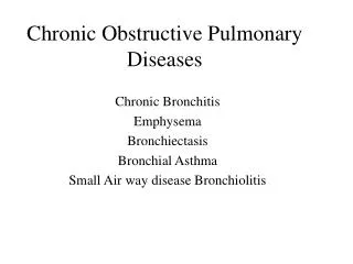
OBSTRUCTIVE AIRWAYS DISEASE • Chronic bronchitis • Chronic bronchiolitis (Small Airways Disease) • Emphysema • Bronchiectasis • Bronchial asthma
OBSTRUCTIVE AIRWAYS DISEASE All characterized by airflow limitation, but involve different mechanisms and parts of the respiratory tree • Chronic bronchitis - hypersecretory • Chronic bronchiolitis- obstructive • Emphysema - destructive NB Cigarette smoking Frequently co-exist – but 2 clinical syndromes “Blue bloater vs. Pink puffer”
OBSTRUCTIVE AIRWAYS DISEASE – Chronic Bronchitis “Persistent or recurrent excess of secretion in the bronchial tree on most days for at least 3 months in the year, over 2 years” • Middle-aged & elderly, M > F • Mucoid sputum – H. Inf, Strep pneum., Bran. Cat • Cigarette smoke, air pollution, dust exposure – cadmium, smog • At PM - bronchi filled with mucous / pus • Enlargement of submucosal glands (Reid Index) shift to pure mucous from mixed sero-mucinous type • Inceased nos of goblet cells in epithelium, at expense of ciliated cells and Clara cells
OBSTRUCTIVE AIRWAYS DISEASE Acute on chronic bronchitis
OBSTRUCTIVE AIRWAYS DISEASE Loss of airway ‘tapering’ in chronic bronchitis
OBSTRUCTIVE AIRWAYS DISEASE – Small Airway Disease • Airways < 2mm = small bronchi, proximal bronchioles • Bronchiolar goblet cell metaplasia – loss of clara cells – loss of protease inhibitor • Chronic inflammation & fibrosis – focal stenoses • Hypoxic pulmonary vasoconstriction – hypertension – cor pulmonale • Compensatory polycythaemia
OBSTRUCTIVE AIRWAYS DISEASE – Emphysema • Emphysema is a condition of the lung characterized by abnormal, permanent enlargement of the airspaces distal to the terminal bronchioles, accompanied by destruction of their walls and without obvious fibrosis • Airflow limitation is due to premature closure of airways because of diminished elastic recoil
OBSTRUCTIVE AIRWAYS DISEASE – Emphysema Morphologic types according to part of acinus affected • Centriacinar – cigs, UL • Panacinar - 1-AT defficiency, LL • Paraseptal - septal / subpleural
OBSTRUCTIVE AIRWAYS DISEASE CENTRILOBULAR EMPHYSEMA SEPTAL EMPHYSEMA
OBSTRUCTIVE AIRWAYS DISEASE PARASEPTAL EMPHYSEMA Large solitary bullae These may grow large enough to cause respiratory failure by compressing adjacent ‘normal’ lung. Corrective bullectomy or ‘lung reduction’ may return pulmonary function to normal
OBSTRUCTIVE AIRWAYS DISEASE PANACINAR EMPHYSEMA
OBSTRUCTIVE AIRWAYS DISEASE PANACINAREMPHYSEMA
OBSTRUCTIVE AIRWAYS DISEASE – Emphysema - pathogenesis Proteases (elastase) vs. Antiproteases • Neutrophils & macrophages - sources of elastase – increased in smokers / infection / inflamm • Smoking stimulates release and enhances activity of elastase • Oxidants in cig smoke inhibit native 1-AT activity • 1-AT defficiency - unopposed elastase activity • 1-AT specified by proteinase inhibitor (Pi) locus – chrom 14, polymorphism – 70 different variants PiMM – normal, Z and S mutants NB medical relevance
OBSTRUCTIVE AIRWAYS DISEASE – Bronchial Asthma • Asthma - characterized by hyperreactive airways leading to episodic, reversible bronchconstriction, owing to increased responsiveness of the tracheobronchial tree to various stimuli • Extrinsic / Atopic / Allergic = allergy to exogenous substances • Intrinsic / idiosyncratic / Non-atopic = no exogenous factors identified
OBSTRUCTIVE AIRWAYS DISEASE – AtopicAsthma • Commoner • Childhood, M>F • Less severe as age – but 30% symptoms as adults • Assoc eczema, rhinitis • Environmental triggers • Type I (IgE-mediated) hypersensitivity reaction
OBSTRUCTIVE AIRWAYS DISEASE – Nonatopic Asthma • Adult onset • Chronic, tending to worsen with age • Triggered by respiratory tract infxn – viral • Family hx – uncommon • Serum IgE – normal • Virus-induced inflammation may lower threshold of receptors to irritants
OBSTRUCTIVE AIRWAYS DISEASE – Asthma • Sputum – yellow – MPO • Eosinophils, Charcot-Leyden crystals, Curschmann’s spirals and Creola bodies • Lungs at PM – Status Asthmaticus – overdistension, mucous plugging • Micro – luminal mucous & eo, goblet cell hyperplasia, infiltration by eosinophils, BM thickening, bronchial smooth muscle hyperplasia, hypertrophy
(a)Curschmann spiral OBSTRUCTIVE AIRWAYS DISEASE (b)Creolabody b a
OBSTRUCTIVE AIRWAYS DISEASE Hyperinflated lungs in status asthmaticus
OBSTRUCTIVE AIRWAYS DISEASE Sticky mucus plugs in status asthmaticus
“SMOKING-ASSOCIATED” INTERSTITIAL LUNG DISEASE • Respiratory-bronchiolitis (RB) • Desquamative interstitial pneumonia (DIP) • Langerhan’s cell histiocytosis (LCH) Eosinophilic granuloma (EG) Histiocytosis X (HX)
RESPIRATORY (SMOKERS) BRONCHIOLITIS (RB) and DIP • Cough & dyspnoea • LL interstitial infiltrates, restrictive PFTs • Patchy disease • Accumulation of macrophages containing yellow-brown pigment in lumens of distal bronchioles, alveolar ducts & spaces • Mild interstitial thickening • DIP – diffuse filling of alveolar spaces • ?different ends of the spectrum of one disease
LANGERHAN’S CELL HISTIOCYTOSIS (LCH) EOSINOPHILIC GRANULOMA (EG) HISTIOCYTOSIS X (HX) • Pulmonary LCH – smokers • Cough, dyspnoea, fever, malaise, spontaneous pneumothorax • Imaging – UL, cysts and nodules • Micro: discrete stellate nodules, bronchocentric • Langerhan’s cells, histiocytes, eosinophils, • Langerhan’s cells – large histiocytes – “groovy” nuclei • Cysts, stellate or starfish-shaped scars • H&E diagnosis, IHC has replaced EM as a diagnostic tool