Download
1 / 91
1.43k likes | 2.77k Vues
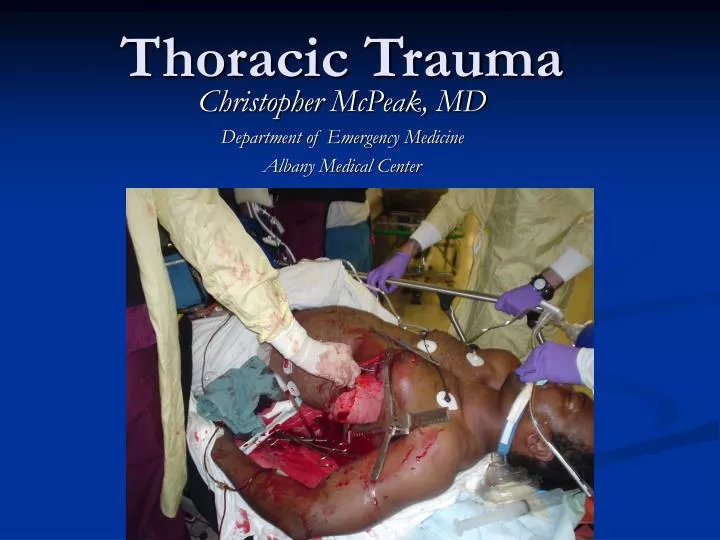
Thoracic Trauma. Christopher McPeak, MD Department of Emergency Medicine Albany Medical Center. Thoracic Trauma. Second leading cause of trauma deaths after head injury Cause of about 10-20% of all trauma deaths Many deaths due to thoracic trauma are preventable. Thoracic Trauma.

E N D
Thoracic Trauma Christopher McPeak, MD Department of Emergency Medicine Albany Medical Center
Thoracic Trauma • Second leading cause of trauma deaths after head injury • Cause of about 10-20% of all trauma deaths • Many deaths due to thoracic trauma are preventable
Thoracic Trauma • Prevention Strategies • Gun Safety Education • Sports Training & Protective Equipment • Seat Belt & Air Bag Use
Thoracic Trauma • Mechanisms of Injury • Blunt Injury • Deceleration • Compression • Penetrating Injury
Thoracic Trauma • Anatomical Injuries • Thoracic Cage (Skeletal) • Cardiovascular • Pleural and Pulmonary • Mediastinal • Diaphragmatic • Esophageal • Penetrating Cardiac
Thoracic Trauma • Often result in: • Hypoxia • Hypercarbia • Acidosis • hypoperfusion of tissues (metabolic)
Thoracic Trauma--Cardiac Impairments to cardiac output • blood loss • increased intrapleural pressures • blood in pericardial sac • myocardial valve damage • vascular disruption
Thoracic Trauma--Respiratory Impairments in ventilatory efficiency • chest excursion compromise • pain • air in pleural space • asymmetrical movement • bleeding in pleural space • ineffective diaphragm contraction
Thoracic Trauma--Respiratory Impairments in gas exchange • atelectasis • pulmonary contusion • respiratory tract disruption
Thoracic Trauma--Exam • Initial exam directed toward life threatening: • Injuries • Open pneumothorax • Flail chest • Tension pneumothorax • Massive hemothorax • Cardiac tamponade • Conditions • Apnea • Respiratory Distress
Thoracic Trauma--Exam • Assessment Findings • Mental Status (decreased) • Pulse (absent, tachy or brady) • BP (narrow PP, hyper- or hypotension, pulsus paradoxus) • Ventilatory rate & effort (tachy- or bradypnea, labored, retractions) • Skin (diaphoresis, pallor, cyanosis, open injury, ecchymosis)
Thoracic Trauma--Exam • Assessment Findings • Neck (tracheal position, SQ emphysema, JVD, open injury) • Chest (contusions, tenderness, asymmetry, absent or decreased lung sounds, bowel sounds, abnormal percussion, open injury, impaled object, crepitus, hemoptysis) • Heart Sounds (muffled, distant, regurgitant murmur) • Upper abdomen (contusion, open injury)
Thoracic Trauma--Exam • Assessment Findings • ECG (ST segment abnormalities, dysrhythmias) • History • Dyspnea • Pain • Past hx of cardiorespiratory disease • Restraint devices used • Item/Weapon involved in injury
Thoracic Trauma Specific Injuries
Rib Fracture • Most common chest wall injury from direct trauma • More common in adults than children • Especially common in elderly • Ribs form rings • Possibility of break in two places • Most commonly 5th - 9th ribs • Poor protection
Rib Fracture • Fractures of 1st and 2nd second require high force • Frequently have injury to aorta or bronchi • Occur in 90% of patients with tracheo-bronchial rupture • May injure subclavian artery/vein • May result in pneumothorax • 30% will die
Rib Fracture • Fractures of 10 to 12th ribs can cause damage to underlying abdominal solid organs: • Liver • Spleen • Kidneys
Rib Fracture • Assessment Findings • Localized pain, tenderness • Increases on palpation or when patient: • Coughs • Moves • Breathes deeply • “Splinted” Respirations • Instability in chest wall, Crepitus • Deformity and discoloration • Associated pneumo or hemothorax
Rib Fracture • Management • High concentration O2 • Positive pressure ventilation as needed • Splint using pillow or swathes • Encourage pt to breath deeply • Helps prevent atelectasis • Analgesics for isolated trauma • Non-circumferential splinting
Rib Fracture • Management • Monitor elderly and COPD patients closely • Broken ribs can cause decompensation • Patients will fail to breathe deeply and cough, resulting in poor clearance of secretions • Usually Non-Emergent Transport
Sternal Fracture • Uncommon, 5-8% in blunt chest trauma • Large traumatic force • Direct blow to front of chest by • Deceleration • steering wheel • dashboard • Other object
Sternal Fracture • 25 - 45% mortality due to associated trauma: • Disruption of thoracic aorta • Tracheal or bronchial tear • Diaphragm rupture • Flail chest • Myocardial trauma • High incidence of myocardial contusion, cardiac tamponade or pulmonary contusion
Sternal Fracture • Assessment Findings • Localized pain • Tenderness over sternum • Crepitus • Tachypnea, Dyspnea • ECG changes with associated myocardial contusion • Hx/Mechanism of blunt chest trauma
Sternal Fracture • Management • Establish airway • High concentration oxygen • Assist ventilations with BVM as needed • IV NS/LR • Emergent Transport • Trauma center
Flail Chest Two or more adjacent ribs fractured in two or more places producing a free floating segment of the chest wall
Flail Chest • Usually secondary to blunt trauma • Most commonly in MVC • Also results from • falls from heights • industrial accidents • assault • birth trauma • More common in older patients
Flail Chest • Mortality rates 20-40% due to associated injuries • Mortality increased with • advanced age • seven or more rib fractures • three or more associated injuries • shock • head injuries
Flail Chest • Consequences of flail chest • Respiratory failure due to • pulmonary contusion • intrathoracic injury • inadequate diaphragm movement • Paradoxical movement of the chest • must be large to compromise ventilation • Increased work of breathing • Pain, decreased chest expansion • leading decreased ventilation
Flail Chest • Consequences of flail chest • Contusion of lung • decreased lung compliance • intra alveolar-capillary hemorrhage • Decreased ventilation • Hypercapnea • Hypoxia
Flail Chest • Assessment Findings • Chest wall contusion • Respiratory distress • Pleuritic chest pain • Splinting of affected side • Crepitus • Tachypnea, Tachycardia • Paradoxical movement (possible)
Flail Chest • Management • Suspect spinal injuries • Establish airway • High concentration oxygen • Assist ventilation with BVM • Treat hypoxia from underlying contusion • Promote full lung expansion • Consider need for intubation and PEEP • Mechanically stabilize chest wall • questionable value
Flail Chest • Management • IV of LR/NS • Avoid rapid replacement in hemodynamically stable patient • Contused lung cannot handle fluid load • Monitor EKG • Chest trauma can cause dysrhythmias • Emergent Transport • Trauma center
Simple Pneumothorax • Incidence • 10-30% in blunt chest trauma • almost 100% with penetrating chest trauma • Morbidity & Mortality dependent on • extent of atelectasis • associated injuries
Simple Pneumothorax • Causes • Commonly a fx rib lacerates lung • May occur spontaneously in tall, thin young males following: • Exertion • Coughing • Air Travel
Simple Pneumothorax • Pathophysiology • Air enters pleural space causing partial lung collapse • small tears self-seal • larger tears may progress • Usually well-tolerated in the young & healthy • Severe compromise can occur in the elderly or patients with pulmonary disease • Degree of distress depends on amount and speed of collapse
Simple Pneumothorax • Assessment Findings • Tachypnea, Tachycardia • Difficulty breathing or respiratory distress • Pleuritic pain • may be referred to shoulder or arm on affected side • Decreased or absent breath sounds • not always reliable • if patient standing, assess apices first • if supine, assess anteriorly • patients with multiple ribs fractures may splint injured side by not breathing deeply
Simple Pneumothorax • Management • Establish airway • High concentration O2 with NRB • Assist with BVM • IV of LR/NS • Monitor for progression • Monitor ECG • Usually Non-emergent transport
Open Pneumothorax Hole in chest wall that allows air to enter pleural space. Larger the hole the more likely air will enter there than through the trachea.
Open Pneumothorax • If the trauma patient does not ventilate well with an open airway, look for a hole • May be subtle • Abrasion with deep punctures
Open Pneumothorax • Pathophysiology • Result of penetrating trauma • Profound hypoventilation may occur • Allows communication between pleural space and atmosphere • Prevents development of negative intrapleural pressure • Results in ipsilateral lung collapse • inability to ventilate affected lung
Open Pneumothorax • Pathophysiology • V/Q Mismatch • shunting • hypoventilation • hypoxia • large functional dead space • Pressure may build within pleural space • Return from Vena cava may be impaired
Open Pneumothorax • Assessment Findings • Opening in the chest wall • Sucking sound on inhalation • Tachycardia • Tachypnea • Respiratory distress • SQ Emphysema • Decreased lung sounds on affected side
Open Pneumothorax • Management • Cover chest opening with occlusive dressing • High concentration O2 • Assist with positive pressure ventilations prn • Monitor for progression to tension pneumothorax • IV with LR/NS • Monitor ECG • Emergent Transport • Trauma Center
Tension Pneumothorax • Incidence • Penetrating Trauma • Blunt Trauma • Morbidity/Mortality • Severe hypoventilation • Immediate life-threat if not managed early
Tension Pneumothorax • Pathophysiology • One-way valve forms in lung or chest wall • Air enters pleural space, but cannot leave • Air is trapped in pleural space • Pressure collapses lung on affected side • Mediastinal shift to contralateral side • Reduction in cardiac output • Increased intrathoracic pressure • deformed vena cava reducing preload
Tension Pneumothorax • Assessment Findings - Most Likely • Severe dyspnea extreme resp distress • Restlessness, anxiety, agitation • Decreased/absent breath sounds • Worsening or Severe Shock / Cardiovascular collapse • Tachycardia • Weak pulse • Hypotension • Narrow pulse pressure
Tension Pneumothorax • Assessment Findings - Less Likely • Jugular Vein Distension • absent if also hypovolemic • Hyperresonance to percussion • Subcutaneous emphysema • Tracheal shift away from injured side (late) • Cyanosis (late)
Tension Pneumothorax • Management • Recognize & Manage early • Establish airway • High concentration O2 • Positive pressure ventilations w/BVM prn • Needle thoracostomy • IV of LR/NS • Monitor ECG • Emergent Transport • Consider need to intubate • Trauma Center preferred
Tension Pneumothorax • Management • Needle Thoracostomy Review • Decompress with 14g (lg bore), 2-inch needle • Midclavicular line: 2nd intercostal space • Midaxillary line: 4-5th intercostal space • Go over superior margin of rib to avoid blood vessels • Be careful not to kink or bend needle or catheter • If available, attach a one-way valve
Hemothorax • Pathophysiology • Blood in the pleural space • Most common result of major trauma to the chest wall • Present in 70 - 80% of penetrating and major non-penetrating trauma cases • Associated with pneumothorax • Rib fractures are frequent cause