Download
1 / 48
490 likes | 816 Vues
Thoracic Trauma. Temple College EMS Professions. Chest Trauma. Second leading cause of trauma deaths after head injury About 20% of all trauma deaths. Chest Trauma. Initial exam directed toward: Open pneumothorax Flail chest Tension pneumothorax Massive hemothorax Cardiac tamponade.

E N D
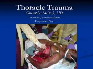
Thoracic Trauma Temple College EMS Professions
Chest Trauma • Second leading cause of trauma deaths after head injury • About 20% of all trauma deaths
Chest Trauma • Initial exam directed toward: • Open pneumothorax • Flail chest • Tension pneumothorax • Massive hemothorax • Cardiac tamponade
Rib Fracture • Most common chest injury • More common in adults than children • Especially common in elderly • Ribs form rings • Consider possibility of break in two places
Rib Fracture • Most commonly 5th to 9th ribs • Poor protection
Rib Fracture • Fractures of 1st, 2nd ribs require high force • Frequently have injury to aorta or bronchi • 30% will die
Rib Fracture • Fractures of 8th to 12th ribs can damage underlying abdominal solid organs: • Liver • Spleen • Kidneys
Rib Fracture • Signs and Symptoms • Localized pain, tenderness • Increases when patient: • Coughs • Moves • Breathes deeply • Chest wall instability • Deformity, discoloration • Associated pneumo or hemothorax
Rib Fracture • Management • High concentration O2 • Splint using pillow, swathes • Encourage patient to breath deeply
Rib Fracture • Management • Monitor elderly and COPD patients carefully • Broken ribs can cause decompensation • Patients will fail to breath deeply and cough, resulting in poor clearance of secretions
Flail Chest • Two or more adjacent ribs broken in two or more places • Produces free-floating chest wall segment • Usually secondary to blunt trauma • More common in older patients
Flail Chest • Signs and Symptoms • Paradoxical movement • May NOT be present initially due to intercostal muscle spasms • Be suspicious in any patient with chest wall: • Tenderness • Crepitus
Flail Chest • Consequences • Pain, leading to decreased ventilation • Increased work of breathing • Contusion of lung
Flail Chest • Management • Establish airway • Suspect spinal injuries • Assist ventilation with BVM and oxygen • Stabilize chest wall
Simple Pneumothorax • Air in pleural space • Partial or complete lung collapse occurs
Simple Pneumothorax • Causes • Chest wall penetration • Fractured rib lacerating lung • Paper bag effect • May occur spontaneously following: • Exertion • Coughing • Air Travel
Simple Pneumothorax • Signs and Symptoms • Pain on inhalation • Difficulty breathing • Tachypnea • Decreased or absent breath sounds Severity of symptoms depends on size of pneumothorax, speed of lung collapse, and patient’s health status
Simple Pneumothorax • Management • Establish airway • Suspect spinal injury based on mechanism • High concentration O2 with NRB • Assist decreased or rapid respirations with BVM • Monitor for tension pneumothorax
Open Pneumothorax • Hole in chest wall • Allows air to enter pleural space • Larger hole = Greater chance air will enter there than through trachea “Sucking Chest Wound”
Open Pneumothorax • Management • Close hole with occlusive dressing • High concentration O2 • Assist ventilations • Consider transport on injured side • Monitor for tension pneumothorax
Tension Pneumothorax • One-way valve forms in lung or chest wall • Air enters pleural space; cannot leave • Air is trapped in pleural space • Pressure rises • Pressure collapses lung
Tension Pneumothorax • Trapped air pushes heart, lungs awayfrom injured side • Vena cavae become kinked • Blood cannot return to heart • Cardiac output falls
Tension Pneumothorax • Signs and Symptoms • Extreme dyspnea • Restlessness, anxiety, agitation • Decreased breath sounds • Hyperresonance to percussion • Cyanosis • Subcutaneous emphysema
Tension Pneumothorax • Signs and Symptoms • Rapid, weak pulse • Decreased BP • Tracheal shift away from injured side • Jugular vein distension Early dyspnea/hypoxia - Late shock
Tension Pneumothorax • Management • Secure airway • High concentration O2 with NRB • If available, request ALS intercept for pleural decompression
Hemothorax • Blood in pleura space • Most common result of major chest wall trauma • Present in 70 to 80% of penetrating, major non-penetrating chest trauma
Hemothorax • Signs and Symptoms • Rapid, weak pulse • Cool, clammy skin • Restlessness, anxiety • Thirst • Chills • Hypotension • Collapsed neck veins
Hemothorax • Signs and Symptoms • Decreased breath sounds • Dullness to percussion • Dyspnea • Ventilatory failure Shock precedes ventilatory failure
Hemothorax • Management • Secure airway • Assist breathing with high concentration O2 • Rapid transport
Traumatic Asphyxia • Blunt force to chest causes • Increased intrathoracic pressure • Backward flow of blood out of heart into vessels of upper chest, neck, head
Traumatic Asphyxia • Signs and Symptoms • Possible sternal fracture or central flail chest • Shock • Purplish-red discoloration of: • Head • Neck • Shoulders • Blood shot, protruding eyes • Swollen, cyanotic lips
Traumatic Asphyxia Name given because patients looked like they had been strangled or hanged
Traumatic Asphyxia • Management • Airway with C-spine control • Assist ventilations with high concentration O2 • Spinal stabilization • Rapid transport
Cardiovascular Trauma Any patient with significant blunt or penetrating trauma to chest has heart/great vessel injury until proven otherwise
Myocardial Contusion • Bruise of heart muscle • Most common blunt cardiac injury • Usually due to steering wheel impact
Myocardial Contusion • Behaves like acute MI • May produce arrhythmias • May cause cardiogenic shock, hypotension
Myocardial Contusion • Signs and Symptoms • Cardiac arrhythmias after blunt chest trauma • Angina-like pain unresponsive to nitroglycerin • Chest pain independent of respiratory movement Suspect in all blunt chest trauma
Myocardial Contusion • Management • High concentration O2 • Transport • Consider ALS intercept
Cardiac Tamponade • Rapid accumulation of blood in space between heart, pericardium • Heart compressed • Blood entering heart decreases • Cardiac output falls
Cardiac Tamponade • Signs and Symptoms • Hypotension unresponsive to treatment • Increased central venous pressure (distended neck/arm veins in presence of decreased arterial BP) • Small quiet heart (decreased heart sounds) Beck’s Triad
Cardiac Tamponade • Signs and Symptoms • Narrowing pulse pressure • Pulsus paradoxicus • Radial pulse becomes weak or disappears when patient inhales
Cardiac Tamponade • Management • Secure airway • High concentration O2 • Rapid transport • Definitive treatment is pericardiocentesis followed by surgery
Traumatic Aortic Aneurysm • Caused by sudden decelerations, massive blunt force: • Vehicle collisions • Falls from heights • Crushing chest trauma • Blunt chest trauma • Animal kicks
Traumatic Aortic Aneurysm • Rupture usually occurs just beyond left subclavian artery • Attachment of aorta to pulmonary artery at this point produces shearing force on aortic arch
Traumatic Aortic Aneurysm • Signs and Symptoms • Increased BP in arms in absence of head injury • Decreased femoral pulses with full arm pulses • Respiratory distress • Ache in chest, shoulders, lower back, abdomen. (Only 25% of patients) Detection requires high index of suspicion
Traumatic Aortic Aneurysm • Management • High concentration oxygen • Assist ventilation • Suspect spinal injury • Rapid transport
Associated Abdominal Trauma • Diaphragm forms dome that extends up into rib cage • Trauma to chest below 4th rib = Abdominal injury until proven otherwise
PowerPoint Source • Slides for this presentation from Temple College EMS: http://www.templejc.edu/dept/ems/pages/powerpoint.html