Download
1 / 1
10 likes | 82 Vues
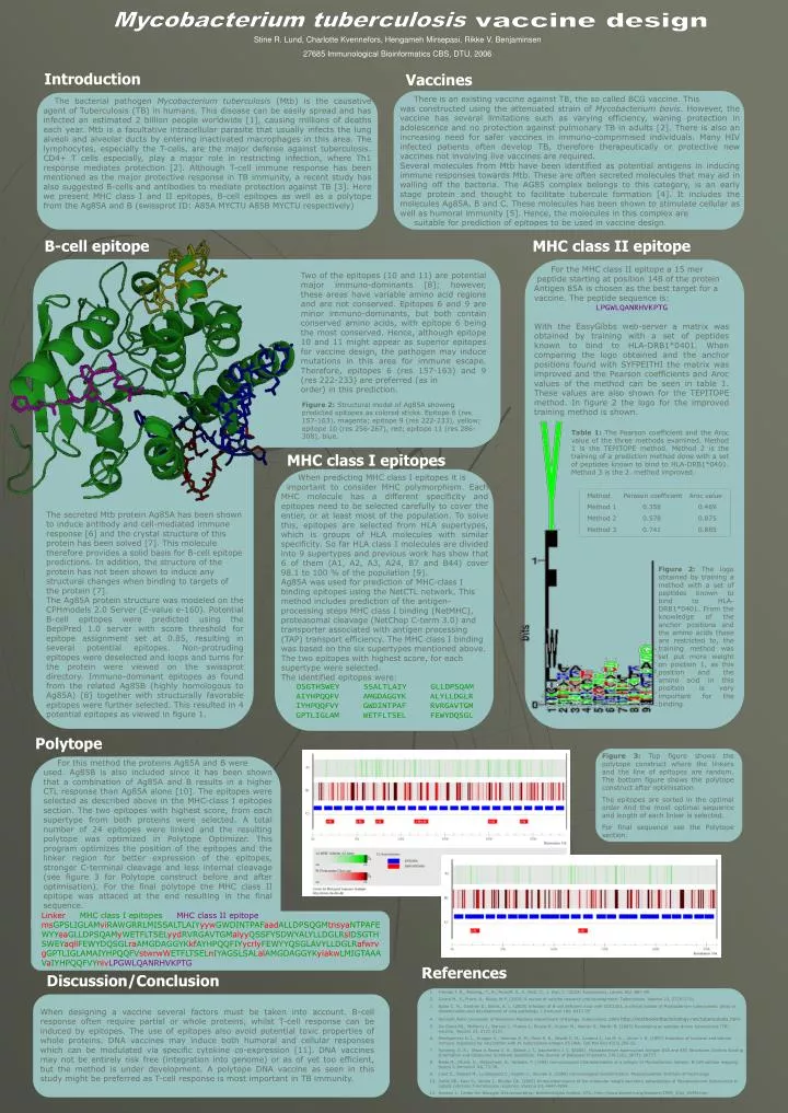
Figure 3: Top figure shows the polytope construct where the linkers and the line of epitopes are random. The bottom figure shows the polytope construct after optimisation. The epitopes are sorted in the optimal order And the most optimal sequence and length of each linker is selected.

E N D
Figure 3: Top figure shows the polytope construct where the linkers and the line of epitopes are random. The bottom figure shows the polytope construct after optimisation. The epitopes are sorted in the optimal order And the most optimal sequence and length of each linker is selected. For final sequence see the Polytope section. Mycobacterium tuberculosis vaccine design Stine R. Lund, Charlotte Kvennefors, Hengameh Mirsepasi, Rikke V. Benjaminsen 27685 Immunological Bioinformatics CBS, DTU, 2006 Introduction Vaccines There is an existing vaccine against TB, the so called BCG vaccine. This was constructed using the attenuated strain of Mycobacterium bovis. However, the vaccine has several limitations such as varying efficiency, waning protection in adolescence and no protection against pulmonary TB in adults [2]. There is also an increasing need for safer vaccines in immuno-comprimised individuals. Many HIV infected patients often develop TB, therefore therapeutically or protective new vaccines not involving live vaccines are required. Several molecules from Mtb have been identified as potential antigens in inducing immune responses towards Mtb. These are often secreted molecules that may aid in walling off the bacteria. The AG85 complex belongs to this category, is an early stage protein and thought to facilitate tubercule formation [4]. It includes the molecules Ag85A, B and C. These molecules has been shown to stimulate cellular as well ashumoral immunity [5]. Hence, the molecules in this complex are suitable for prediction of epitopes to be used in vaccine design. The bacterial pathogen Mycobacterium tuberculosis (Mtb) is the causative agent of Tuberculosis (TB) in humans. This disease can be easily spread and has infected an estimated 2 billion people worldwide [1], causing millions of deaths each year. Mtb is a facultative intracellular parasite that usually infects the lung alveoli and alveolar ducts by entering inactivated macrophages in this area. The lymphocytes, especially the T-cells, are the major defense against tuberculosis. CD4+ T cells especially, play a major role in restricting infection, where Th1 response mediates protection [2]. Although T-cell immune response has been mentioned as the major protective response in TB immunity, a recent study has also suggested B-cells and antibodies to mediate protection against TB [3]. Here we present MHC class I and II epitopes, B-cell epitopes as well as a polytope from the Ag85A and B (swissprot ID: A85A MYCTU A85B MYCTU respectively) B-cell epitope MHC class II epitope For the MHC class II epitope a 15 mer peptide starting at position 148 of the protein Antigen 85A is chosen as the best target for a vaccine. The peptide sequence is: LPGWLQANRHVKPTG With the EasyGibbs web-server a matrix was obtained by training with a set of peptides known to bind to HLA-DRB1*0401. When comparing the logo obtained and the anchor positions found with SYFPEITHI the matrix was improved and the Pearson coefficients and Aroc values of the method can be seen in table 1. These values are also shown for the TEPITOPE method. In figure 2 the logo for the improved training method is shown. Two of the epitopes (10 and 11) are potential major immuno-dominants [8]; however, these areas have variable amino acid regions and are not conserved. Epitopes 6 and 9 are minor immuno-dominants, but both contain conserved amino acids, with epitope 6 being the most conserved. Hence, although epitope 10 and 11 might appear as superior epitopes for vaccine design, the pathogen may induce mutations in this area for immune escape. Therefore, epitopes 6 (res 157-163) and 9 (res 222-233) are preferred (as in order) in this prediction. Figure 2: Structural model of Ag85A showing predicted epitopes as colored sticks. Epitope 6 (res 157-163), magenta; epitope 9 (res 222-233), yellow; epitope 10 (res 256-267), red; epitope 11 (res 286-308), blue. Table 1: The Pearson coefficient and the Aroc value of the three methods examined. Method 1 is the TEPITOPE method. Method 2 is the training of a prediction method done with a set of peptides known to bind to HLA-DRB1*0401. Method 3 is the 2. method improved. MHC class I epitopes When predicting MHC class I epitopes it is important to consider MHC polymorphism. Each MHC molecule has a different specificity and epitopes need to be selected carefully to cover the entier, or at least most of the population. To solve this, epitopes are selected from HLA supertypes, which is groups of HLA molecules with similar specificity. So far HLA class I molecules are divided into 9 supertypes and previous work has show that 6 of them (A1, A2, A3, A24, B7 and B44) cover 98.1 to 100 % of the population [9]. Ag85A was used for prediction of MHC-class I binding epitopes using the NetCTL network. This method includes prediction of the antigen-processing steps MHC class I binding (NetMHC), proteasomal cleavage (NetChop C-term 3.0) and transporter associated with antigen processing (TAP) transport efficiency. The MHC class I binding was based on the six supertypes mentioned above. The two epitopes with highest score, for each supertype were selected. The identified epitopes were: DSGTHSWEY SSALTLAIY GLLDPSQAM AIYHPQQFV AMGDAGGYK ALYLLDGLR IYHPQQFVY GWDINTPAF RVRGAVTGM GPTLIGLAM WETFLTSEL FEWYDQSGL Method Perason coefficient Aroc value Method 1 0.358 0.469 Method 2 0.578 0.875 Method 3 0.741 0.885 The secreted Mtb protein Ag85A has been shown to induce antibody and cell-mediated immune response [6] and the crystal structure of this protein has been solved [7]. This molecule therefore provides a solid basis for B-cell epitope predictions. In addition, the structure of the protein has not been shown to induce any structural changes when binding to targets of the protein [7]. The Ag85A protein structure was modeled on the CPHmodels 2.0 Server (E-value e-160). Potential B-cell epitopes were predicted using the BepiPred 1.0 server with score threshold for epitope assignment set at 0.85, resulting in several potential epitopes. Non-protruding epitopes were deselected and loops and turns for the protein were viewed on the swissprot directory. Immuno-dominant epitopes as found from the related Ag85B (highly homologous to Ag85A) [8] together with structurally favorable epitopes were further selected. This resulted in 4 potential epitopes as viewed in figure 1. Figure 2: The logo obtained by training a method with a set of peptides known to bind to HLA-DRB1*0401. From the knowledge of the anchor positions and the amino acids these are restricted to, the training method was set put more weight on position 1, as this position and the amino acid in this position is very important for the binding. Polytope For this method the proteins Ag85A and B were used. Ag85B is also included since it has been shown that a combination of Ag85A and B results in a higher CTL response than Ag85A alone [10]. The epitopes were selected as described above in the MHC-class I epitopes section. The two epitopes with highest score, from each supertype from both proteins were selected. A total number of 24 epitopes were linked and the resulting polytope was optimized in Polytope Optimizer. This program optimizes the position of the epitopes and the linker region for better expression of the epitopes, stronger C-terminal cleavage and less internal cleavage (see figure 3 for Polytope construct before and after optimisation). For the final polytope the MHC class II epitope was attaced at the end resulting in the final sequence. Linker MHC class I epitopes MHC class II epitope msGPSLIGLAMviRAWGRRLMISSALTLAIYyywGWDINTPAFaadALLDPSQGMtnsyaNTPAFEWYYeaGLLDPSQAMyWETFLTSELyydRVRGAVTGMalyyQSSFYSDWYALYLLDGLRslDSGTHSWEYaqllFEWYDQSGLraAMGDAGGYKkfAYHPQQFIYycrlyFEWYYQSGLAVYLLDGLRafwrvgGPTLIGLAMAIYHPQQFVstwrwWETFLTSELnIYAGSLSALalAMGDAGGYKyiakwLMIGTAAAVaIYHPQQFVYnivLPGWLQANRHVKPTG References Discussion/Conclusion • Frieden T. R., Sterling., T., R., Munsiff, S., S. Watt, C., J. Dye, C. (2003) Tuberculosis, Lancet 362, 887-99. • Girard M., P., Fruth, U., Kieny, M-P. (2005) A review of vaccine research and development: Tuberculosis. Vaccine 23, 2725-2731. • Bosio C. M., Gardner D., Elkins, K. L. (2000) Infection of B-cell deficient mice with CDC1551, a clinical isolate of Myobacterium tuberculosis: delay in dissemination and development of lung pathology. J Immunol 164, 6417-25 • Kenneth Todar University of Wisconsin-Madison Department of Biology. Tuberculosis. 2005 http://textbookofbacteriology.net/tuberculosis.html • De Groot AS., McMurry J., Marcon L., Franco J., Rivera D., Kutzler M., Weiner D., Martin B. (2005) Developing an epitope-driven tuberculosis (TB) vaccine. Vaccine 23, 2121-2131. • Montgomery D. L., Huygen K., Yawman A. M., Deck R. R., Dewitt C. M., Content J., Liu M. A. , Ulmer J. B. (1997) Induction of humoral and cellular immune responses by vaccination with M. tuberculosis antigen 85 DNA. Cell Mol Biol 43(3),285-92. • Ronning, D. R., Vissa V, Besra G. S., Belisle J. T., Sacchettini J. C. (2004).Mycobacterium tuberculosis Antigen 85A and 85C Structures Confirm Binding Orientation and Conserved Substrate Specificity. The Journal of Biological Chemistry 279 (35), 36771-36777. • Naito M., Ohara, O., Matsumoto, S., Yamada, T. (1998) Immunological Characterization of α antigen of Myobacterium kansaii: B-Cell epitope mapping. Scand J. Immunol. 48, 73-78. • Lund C., Nielsen M., Lundegaard C., Keşmir C., Brunak S. (2005) Immunological bioinformatics. Massachusettes Institute of Technology. • Sable SB., Kaur S., Verma I., Khuller GK. (2005) Immunodominance of low molecular weight secretory polupeptides of Mycobacterium tuberculosis to induce cytotoxic T-lymphocyte response. Vaccine 23, 4947-4954. • Hansen J., Center for Biologisk Sekvensanalyse, Bioteknologisk Institut, DTU. http://www.biokemi.org/biozoom/1999_3/bz_0399f.htm When designing a vaccine several factors must be taken into account. B-cell response often require partial or whole proteins, whilst T-cell response can be induced by epitopes. The use of epitopes also avoid potential toxic properties of whole proteins. DNA vaccines may induce both humoral and cellular responses which can be modulated via specific cytokine co-expression [11]. DNA vaccines may not be entirely risk free (integration into genome) or as of yet too efficient, but the method is under development. A polytope DNA vaccine as seen in this study might be preferred as T-cell response is most important in TB immunity.