Download
1 / 37
380 likes | 758 Vues
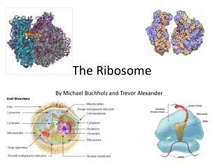
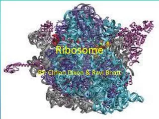
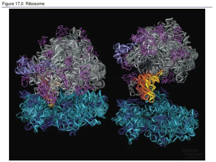
Figure 17.0 Ribosome. Figure 17.1 Beadle and Tatum’s evidence for the one gene-one enzyme hypothesis. Figure 17.2 Overview: the roles of transcription and translation in the flow of genetic information (Layer 1).

E N D
Figure 17.1 Beadle and Tatum’s evidence for the one gene-one enzyme hypothesis
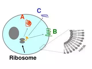
Figure 17.2 Overview: the roles of transcription and translation in the flow of genetic information (Layer 1)
Figure 17.2 Overview: the roles of transcription and translation in the flow of genetic information (Layer 2)
Figure 17.2 Overview: the roles of transcription and translation in the flow of genetic information (Layer 3)
Figure 17.2 Overview: the roles of transcription and translation in the flow of genetic information (Layer 4)
Figure 17.2 Overview: the roles of transcription and translation in the flow of genetic information (Layer 5)
Figure 17.6 The stages of transcription: initiation, elongation, and termination (Layer 1)
Figure 17.6 The stages of transcription: initiation, elongation, and termination (Layer 2)
Figure 17.6 The stages of transcription: initiation, elongation, and termination (Layer 3)
Figure 17.6 The stages of transcription: initiation, elongation, and termination (Layer 4)
Figure 17.7 The initiation of transcription at a eukaryotic promoter
Figure 17.8 RNA processing; addition of the 5 cap and poly(A) tail
Figure 17.10 The roles of snRNPs and spliceosomes in mRNA splicing
Figure 17.11 Correspondence between exons and protein domains
Figure 17.14 An aminoacyl-tRNA synthetase joins a specific amino acid to a tRNA
Figure 17.16 Structure of the large ribosomal subunit at the atomic level
Figure 17.21 The signal mechanism for targeting proteins to the ER
Figure 17.22 Coupled transcription and translation in bacteria
Figure 17.23 The molecular basis of sickle-cell disease: a point mutation
Figure 17.24 Categories and consequences of point mutations: Base-pair insertion or deletion
Figure 17.24 Categories and consequences of point mutations: Base-pair substitution
Figure 17.25 A summary of transcription and translation in a eukaryotic cell