Download
1 / 25
610 likes | 1.85k Vues
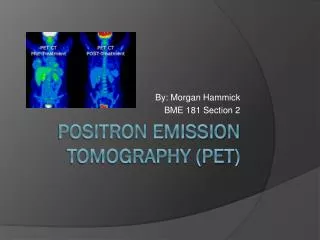
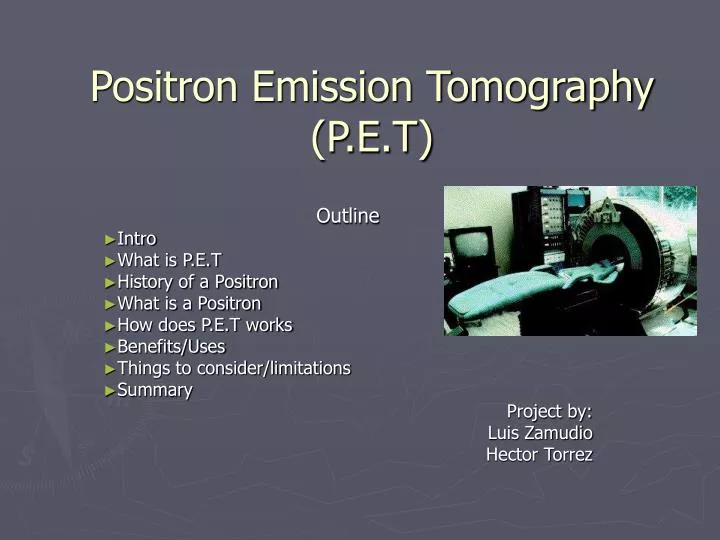
Positron Emission Tomography (P.E.T). Outline Intro What is P.E.T History of a Positron What is a Positron How does P.E.T works Benefits/Uses Things to consider/limitations Summary Project by: Luis Zamudio Hector Torrez. What is PET.

E N D
Positron Emission Tomography (P.E.T) Outline Intro What is P.E.T History of a Positron What is a Positron How does P.E.T works Benefits/Uses Things to consider/limitations Summary Project by: Luis Zamudio Hector Torrez
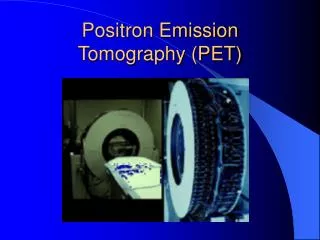
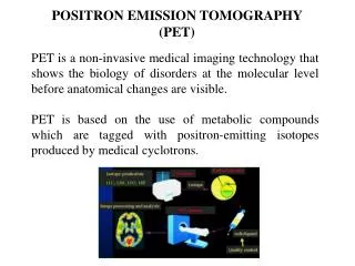
What is PET • PET is a noninvasive, diagnostic imaging technique for measuring the metabolic activity of cells in the human body. • It was developed in the mid 1970s and it was the first scanning method to give functional information about the brain. Htt://www.nucmed.buffalo.edu/petdef.htm
A little history about the positron • Existence first postulated in 1928 by Paul Dirac • First observed in 1932 by Carl D. Anderson, who gave the positron its name. He also suggested to rename the electron to “negatron” but he was unsuccessful. http://en.wikipedia.org/wiki/Positron
What is a Positron? • A Positron is an anti-matter electron, it is identical in mass but has an apposite charge of +1. • Positron can come from different number of sources, but for PET they are produced by nuclear decay. • Nuclear decay is basically when unstable nuclei are produced in a cyclotron by bombarding the target material with protons, and as a result a neutron is released. 18-O + proton => 18-F + neutron • In PET the target material is chosen so that the product of the bombardment decays to a more stable state isotope by emitting a positron, for instance 18-F has too many protons, so one of these protons decays into a neutron emitting in the process a positron an a neutrino. • proton (+1 charge) => neutron (0 charge) + positron (+1 charge) + neutrino (0 charge) • After decay, we’re left with 18-O http://www.nucmed.buffalo.edu/positron.htm
What happens after the positron is obtained? • Left over energy from the nuclear decay process is shared between the positron and the departing neutrino. Kinetic energy. • Because of conservation of energy and momentum the positron is forced to stay and thus become useful. • Positron begins its activity in colliding with other particles and gradually losing its kinetic energy and thus slowing down. http://www.nucmed.buffalo.edu/positron.htm
Annihilation of a positron and electron • The positron will encounter an electron and completely annihilate each other resulting in converting all their masses into energy. This is the result of two photons, or gamma rays. • Because of conservation of energy and momentum, each photon has energy of 511keV and head in an almost 180 degrees from each other. • 511keV is the ideal rest state annihilation value. http://www.nucmed.buffalo.edu/positron.htm
How do we detect photons (gamma rays)? • PET detects these photons with a PET camera which allows to determine where they came from, where the nucleus was when it decayed, and also knowing where the nucleus goes in the body. http://www.nucmed.buffalo.edu/positron.htm
What are some of the uses for PET • Patients with conditions affecting the brain • Heart • Certain types of Cancer • Alzheimer’s disease • Some neurological disorders Htt://www.nucmed.buffalo.edu/petdef.htm
Patients with brain disorders • PET scans of the brain are used to evaluate patients who have memory disorders of an undetermined cause, suspected or proven brain tumors or seizure disorders that are not responsive to medical therapy and are therefore candidates for surgery. http://www.radiologyinfo.org/content/petomography.htm
Heart Conditions • PET scans of the heart are used to determine blood flow to the heart muscle and help evaluate signs of coronary artery disease. PET scans of the heart can also be used to determine if areas of the heart that show decreased function are alive rather than scarred as a result of a prior heart attack, called a myocardial infarction. Combined with a myocardial perfusion study, PET scans allow differentiation of nonfunctioning heart muscle from heart muscle that would benefit from a procedure, such as coronary bypass for instance. http://www.radiologyinfo.org/content/petomography.htm
Cancer Patients • Used to determine if there are new or advancing cancers by analysis of biochemical changes. • It is used to examine the effects of cancer therapy by characterizing biochemical changes in the cancer. PET scans can be performed on the whole body. http://www.radiologyinfo.org/content/petomography.htm
Alzheimer’s disease • With Alzheimer’s disease there is no gross structural abnormality, but PET is able to show a biochemical change.
Neurological disorders • Positron emission tomography (PET) imaging has recently been shown to aid in the diagnosis of particular neurological syndromes associated with cancer. • Before their cancer is even diagnosed, patients can develop problems with the brain, spinal cord or nerves, though the cancer has not spread to the nervous system. Called "paraneoplastic neurological disorders," these neurological problems occur as the body's immune system begins to fight the cancer cells, but accidentally attacks the brain or nerves as well. These problems are uncommon, difficult to diagnose, and usually appear in patients whose primary cancer is extremely difficult to find. Abnormal antibodies in the blood or spinal fluid are often associated with these disorders, though they cannot help identify the primary tumor. http://interactive.snm.org/index.cfm?PageID=2367&RPID=535
How does it work? • Before the examination begins, a radioactive substance is produced in a machine called a cyclotron and attached, or tagged, to a natural body compound, most commonly glucose, but sometimes water or ammonia. Once this substance is administered to the patient, the radioactivity localizes in the appropriate areas of the body and is detected by the PET scanner. • Different colors or degrees of brightness on a PET image represent different levels of tissue or organ function. For example, because healthy tissue uses glucose for energy, it accumulates some of the tagged glucose, which will show up on the PET images. However, cancerous tissue, which uses more glucose than normal tissue, will accumulate more of the substance and appear brighter than normal tissue on the PET images. http://www.radiologyinfo.org/content/petomography.htm
Labeling • Chemical compounds we'd like to follow through the body are labeled with radioactive atoms that decay by emitting positrons. Labeling is a process of attaching some kind of identifying tag to the compound you want to follow which will later let you identify where the compound has gone. In PET the compounds that can be labeled are limited only by the imagination of the investigators and the physical half-life of the positron emitting label. One of the big advantages of PET is that the atoms which can be labeled (turned into positron emitters) are the same atoms which naturally comprise the organic molecules utilized in the body. These atoms include oxygen, carbon and nitrogen to name a few. Since these atoms occur naturally in organic compounds, replacing the naturally occurring atoms in a compound with a labeled atom leaves you a compound that is chemically and biologically identical to the original (so it will behave in a manner identical to its unlabeled sibling) and that is traceable. In addition to naturally occurring compounds such as neurotransmitters, sugars, etc., it is also possible to label synthesized compounds (such as drugs) and follow them as well. http://www.nucmed.buffalo.edu/petdef.htm
Tracers • A second important attribute of PET is that it can follow labeled compounds in trace quantities. This means that the labeled compounds can be introduced into the body without affecting the normal processes of the body. For example, labeling a pound of sugar and ingesting that sugar would be a good example of a non-trace quantity of labeled compound. At these quantities, blood chemistry would be altered (e.g. insulin produced in response to rising blood sugar levels). Often you want to follow the time course of a compound in the body by introducing trace quantities of a compound that will behave the same as the unlabeled compound without altering the ongoing physiological state of chemical processes of the body. PET is sensitive enough to detect trace amounts of labeled compound and so is well suited to this kind of investigation. http://www.nucmed.buffalo.edu/petdef.htm
How is it performed? • A nurse or technologist will take you into a special injection room, where the radioactive substance is administered as an intravenous injection (although in some cases, it will be given through an existing intravenous line or inhaled as a gas). It will then take approximately 30 to 90 minutes for the substance to travel through your body and accumulate in the tissue under study. During this time, you will be asked to rest quietly and avoid significant movement or talking, which may alter the localization of the administered substance. After that time, scanning begins. This may take 30 to 45 minutes. • Some patients, specifically those with heart disease, may undergo a stress test in which PET scans are obtained while they are at rest and again after undergoing the administration of a pharmaceutical to alter the blood flow to the heart. • Usually, there are no restrictions on daily routine after the test, although you should drink plenty of fluids to flush the radioactive substance from your body. http://www.radiologyinfo.org/content/petomography.htm
What are the benefits vs. risks? • Because PET allows study of body function, it can help physicians detect alterations in biochemical processes that suggest disease before changes in anatomy are apparent with other imaging tests, such as CT or MRI. • Because the radioactivity is very short-lived, your radiation exposure is low. The substance amount is so small that it does not affect the normal processes of the body. • PET imaging has been shown to improve detection of a variety of cancers, and earlier tests have suggested this technique may be useful in identifying small tumors in patients with paraneoplastic neurological disorders. • The radioactive substance may expose radiation to the fetus in patients who are pregnant or the infants of women who are breast-feeding. The risk to the fetus or infant should be considered in relation to the potential information gain from the result of the PET examination. If you are pregnant, you should inform the PET imaging staff before the examination is performed. http://www.radiologyinfo.org/content/petomography.htm
Things to consider • You will remain still for a long time. • Claustrophobic persons may feel some anxiety. • Even though you may feel the desire to feel something due to the radioactivity, you will be disappointed, unless they mistakenly inject you plutonium gas.
Limitations • PET can give false results if a patient's chemical balances are not normal. Specifically, test results of diabetic patients or patients who have eaten within a few hours prior to the examination can be adversely affected because of blood sugar or blood insulin levels. • Also, because the radioactive substance decays quickly and is effective for a short period of time, it must be produced in a laboratory near the PET scanner. It is important to be on time for the appointment and to receive the radioactive substance at the scheduled time. PET must be done by a radiologist who has specialized in nuclear medicine and has substantial experience with PET. Most large medical centers now have PET services available to their patients. Medicare and insurance companies cover many of the applications of PET, and coverage continues to increase. • Finally, the value of a PET scan is enhanced when it is part of a larger diagnostic work-up. This often entails comparison of the PET scan with other imaging studies, such as CT or MRI. http://www.radiologyinfo.org/content/petomography.htm
Relevant information • But PET imaging is not yet widely available, and clear indicators of clinically meaningful outcomes using PET are essential to warrant use with this patient population. • "Accurately defining the role of this technique for these patients is critical," comments study author Steven Allder, MD, of the department of Neurology, Royal Hallamshire Hospital in Sheffield, United Kingdom. Toward this end, Adler and colleagues studied the use of PET imaging in 32 patients with suspected paraneoplastic neurological disorders who had not yet been diagnosed with cancer. • With each patient, all relevant investigations had been performed prior to PET imaging resulting in no diagnostic conclusions. Each patient then underwent PET imaging from neck to pelvis. All patients were then prospectively followed-up, with the results of all further investigation collected. Final diagnosis was determined, and the sensitivity and specificity of the results of the initial PET scan were calculated. • "This particular PET scanning in our patient population successfully yielded a high proportion of relevant lesions that were undetectable by alternative diagnostic means," reports Allder. Results of this study indicate that PET is an appropriate, promising tool for patients with undiagnosed paraneoplastic neurological disorders. http://interactive.snm.org/index.cfm?PageID=2367&RPID=535
Summary of P.E.T • PET produces images of the body by detecting the radiation emitted from radioactive substances. These substances are injected into the body, and are usually tagged with a radioactive atom (C-11, Fl-18, O-15 or N-13) that has short decay time. These radioactive atoms are formed by bombarding normal chemicals with neutrons to create short-lived radioactive isotopes. PET detects the gamma rays given off at the site where a positron emitted from the radioactive substance collides with an electron in the tissue. The results are evaluated by a trained expert. http://science.howstuffworks.com/nuclear-medicine2.htm