Download

1 / 102
1.02k likes | 1.04k Vues
The Genetic Code, Mutations, and Translation. OVERVIEW OF TRANSLATION The second stage in gene expression is translating the nucleotide sequence of a messenger RNA molecule into the amino acid sequence of a protein.

E N D
OVERVIEW OF TRANSLATION • The second stage in gene expression is translating the nucleotide sequence of a messenger RNA molecule into the amino acid sequence of a protein. • The genetic code is defined as the relationship between the sequence of nucleotides in DNA (or its RNA transcripts) and the sequence of amino acids in a protein. • Each amino acid is specified by one or more nucleotide triplets (codons) in the DNA. • During translation, mRNA acts as a working copy of the gene in which the codons for each amino acid in the protein have been transcribed from DNA to mRNA.
tRNAs serve as adapter molecules that couple the codons in mRNA with the amino acids they each specify, thus aligning them in the appropriate sequence before peptide bond formation. • Translation takes place on ribosomes, complexes of protein and rRNA that serve as the molecular machines coordinating the interactions between mRNA, tRNA, the enzymes, and the protein factors required for protein synthesis. • Many proteins undergo posttranslational modifications as they prepare to assume their ultimate roles in the cell.
THE GENETIC CODE • Most genetic code tables designate the codons for amino acids as mRNA sequences. Important features of the genetic code include: • Each codon consists of three bases (triplet). There are 64 codons. They are all written in the 5' to 3' direction. • 61 codons code for amino acids. The other three (UAA, UGA, UAG) are stop codons (or nonsense codons) that terminate translation. • There is one start codon (initiation codon), AUG, coding for methionine. Protein synthesis begins with methionine (Met) in eukaryotes, and formylmethionine (fmet) in prokaryotes. • The code is unambiguous. Each codon specifies no more than one amino acid.
The code is degenerate. More than one codon can specify a single amino acid. • All amino acids, except Met and tryptophan (Trp), have more than one codon. • For those amino acids having more than one codon, the first two bases in the codon are usually the same. The base in the third position often varies. • The code is almost universal (the same in all organisms). Some minor exceptions to this occur in mitochondria and some organisms. • The code is commaless (contiguous). There are no spacers or "commas" between codons on an mRNA. • Neighboring codons on a message are non-overlapping.
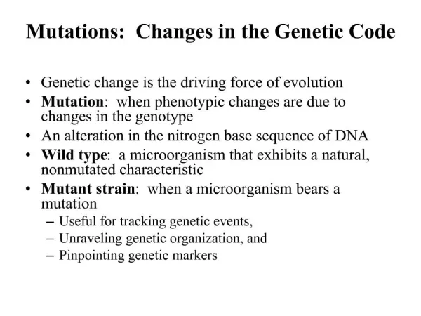
MUTATIONS • A mutation is any permanent, heritable change in the DNA base sequence of an organism. This altered DNA sequence can be reflected by changes in the base sequence of mRNA, and, sometimes, by changes in the amino acid sequence of a protein. • Mutations can cause genetic diseases. They can also cause changes in enzyme activity, nutritional requirements, antibiotic susceptibility, morphology, antigenicity, and many other properties of cells.
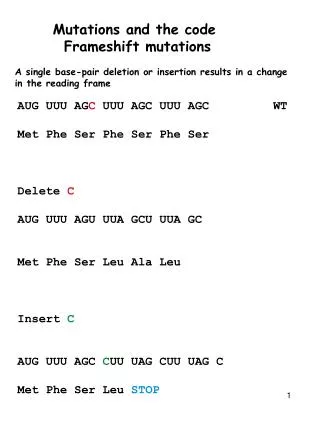
A very common type of mutation is a single base alteration or point mutation. • A transition is a point mutation that replaces a purine-pyrimidine base pair with a different purine-pyrimidine base pair. For example, an A-T base pair becomes a G-C base pair. • A transversion is a point mutation that replaces a purine-pyrimidine base pair with a pyrimidine-purine base pair. For example, an A-T base pair becomes a T-A or a C-G base pair.
Mutations are often classified according to the effect they have on the structure of the gene's protein product. • This change in protein structure can be predicted using the genetic code table in conjunction with the base sequence of DNA or mRNA.
Effect of Some Common Types of Mutations on Protein Structure
Large Segment Deletions • Large segments of DNA can be deleted from a chromosome during an unequal crossover in meiosis. • Crossover or recombination between homologous chromosomes is a normal part of meiosis I that generates genetic diversity in reproductive cells (egg and sperm), a largely beneficial result. • In a normal crossover event, the homologous maternal and paternal chromosomes exchange equivalent segments, and although the resultant chromosomes are mosaics of maternal and paternal alleles, no genetic information has been lost from either one. • On rare occasions, a crossover can be unequal, and one of the two homologs loses some of its genetic information.
α-Thalassemia is a well-known example of a genetic disease in which unequal crossover has deleted one or more α-globin genes from chromosome 16. • Cri-du-chat (mental retardation, microcephaly, wide-set eyes, and a characteristic kitten-like cry) results from a terminal deletion of the short arm of chromosome 5.
Large Segment Deletion During Crossing-Over in Meiosis
Mutations in Splice Sites • Mutations in splice sites affect the accuracy of intron removal from hnRNA during post-transcriptional processing, if a splice site is lost through mutation, spliceosomes may: • Delete nucleotides from the adjacent exon. • Leave nucleotides of the intron in the processed mRNA. • Use the next normal upstream or downstream splice site, deleting an exon from the processed mRNA. • Mutations in splice sites have now been documented in many different diseases including β-thalassemia, Gaucher disease, and Tay-Sachs.
Trinucleotide Repeat Expansion • The mutant alleles in certain diseases, such as Huntington disease, fragile X syndrome, and myotonic dystrophy, differ from their normal counterparts only in the number of tandem copies of a trinucleotide. • The expansion of the trinucleotide repeat in the mutant allele can be in a coding region (Huntington and spinobulbar muscular atrophy) or in an untranslated region of the gene (fragile X and myotonic dystrophy) or even in an intron (Friedrich ataxia). • In these diseases, the number of repeats often increases with successive generations and correlates with increasing severity and decreasing age of onset, a phenomenon called anticipation.
In the normal Huntington allele, there are < 35 tandem repeats of CAG in the coding region. • Affected family members may have > 39 of these CAG repeats. • The normal protein contains five adjacent glutamine residues, whereas the proteins encoded by the disease-associated alleles have 30 or more adjacent glutamines. • The long glutamine tract makes the abnormal proteins extremely unstable.
Clinical Correlate • Huntington's disease, an autosomal dominant disorder, has a mean age-of-onset of 43-48 years. • Symptoms appear gradually and worsen over a period of about 15 years until death occurs. Mood disturbance, impaired memory, and hyperreflexia are often the first signs, followed by abnormal gait, chorea (loss of motor control), dystonia, dementia, and dysphagia. • Cases of juvenile onset (<10 years old) are more severe and most frequently occur when the defective allele is inherited paternally. • About 25% of cases have late onset, slower progression and milder symptoms.
AMINO ACID ACTIVATION AND CODON TRANSLATION BY tRNAs • Inasmuch as amino acids have no direct affinity for mRNA, an adapter molecule, which recognizes an amino acid on one end and its corresponding codon on the other, is required for translation. This adapter molecule is tRNA. Amino Acid Activation • As tRNAs enter the cytoplasm, each combines with its cognate amino acid in a two-step process called amino acid activation.
Each type of amino acid is activated by a different amino acyl tRNA synthetase. • Two high-energy bonds from an ATP are required. • The aminoacyl tRNA synthetase transfers the activated amino acid to the 3' end of the correct tRNA. • The amino acid is linked to its cognate tRNA with an energy-rich bond. • This bond will later supply energy to make a peptide bond linking the amino acid into a protein.
Aminoacyl tRNA synthetases have self-checking functions to prevent incorrectly paired amino acyl tRNAs from forming. • If, however, an aminoacyl tRNA synthetase does release an incorrectly paired product (ala-tRNASer), there is no mechanism during translation to detect the error, and an incorrect amino acid will be introduced into some protein.
Codon Translation by Aminoacyl tRNAs • Each tRNA has an anticodon sequence that allows it to pair with the codon for its cognate amino acid in the mRNA. • Because base pairing is involved, the orientation of this interaction will be complementary and antiparallel. • The arg-tRNAarg has an anticodon sequence, UCG, allowing it to pair with the arginine codon CGA. • The anticodon sequence in tRNA is antiparallel and complementary to the codon translated in mRNA.
Wobble • Many amino acids are specified by more than one codon (redundancy). Frequently, a tRNA can translate more than one of these codons, sparing the cell from making multiple tRNAs to carry the same amino acid. • For instance, the arg-tRNAarg can translate both the CGA and the CGG codons that specify arginine. This phenomenon is known as "Wobble" and can be summarized as follows: • Correct base pairing is required at the first position of the codon (third of anticodon) and the second position of the codon (second of anticodon). • The third position of the codon does not always need to be paired with the anticodon (e.g., it is allowed to "wobble" in some cases).
Modifications in the anticodon affect the pattern ofwobble pairing and therefore are important indetermining tRNA specificity • When bases in the anticodon are modified, further pairing patterns become possible in addition to those predicted by the regular and wobble pairing involving A, C, U, and G
TRANSLATION (PROTEIN SYNTHESIS) • Protein synthesis occurs by peptide bond formation between successive amino acids whose order is specified by a gene and thus by an mRNA. Peptide Bond Formation
During translation, the amino acids are attached to the 3' ends of their respective tRNAs. • The aminoacyl-tRNAs are situated in the P and A sites of the ribosome. • The peptide bond forms between the carboxyl group of the amino acid (or growing peptide) in the P site and the amino group of the next amino acid in the A site. • Proteins are synthesized from the amino to the carboxyl terminus.
Formation of a Peptide Bond by a Ribosome During Translation
Steps of Translation • Translation occurs in the cytoplasm of both prokaryotic (Pr) and eukaryotic (Eu) cells. • In prokaryotes, ribosomes can begin translating the mRNA even before RNA polymerase completes its transcription. • In eukaryotes, translation and transcription are completely separated in time and space with transcription in the nucleus and translation in the cytoplasm. • The process of protein synthesis occurs in three stages: initiation, elongation, and termination. • Special protein factors for initiation (IF), elongation (EF), and termination (release factors), as well as GTP, are required for each of these stages.
Initiation • The small ribosomal subunit binds to the mRNA. In prokaryotes, the 16S rRNA of the small subunit binds to the Shine-Dalgarno sequence in the 5' untranslated region of the mRNA. • In eukaryotes, the small subunit binds to the 5' cap structure and slides down the message to the first AUG. • The charged initiator tRNA becomes bound to the AUG start codon on the message through base pairing with its anticodon. • The initiator tRNA in prokaryotes carries fmet, whereas the initiator tRNA in eukaryotes carries Met.
The large subunit binds to the small subunit, forming the completed initiation complex. • There are two important binding sites on the ribosome called the P site and the A site, a third (E site) has been proposed. • The peptidyl site (P site) is the site on the ribosome where (f)met-tRNAi initially binds. After formation of the first peptide bond, the P site is a binding site for the growing peptide chain. • The aminoacyl site (A site) binds each new incoming tRNA molecule carrying an activated amino acid.
Elongation • Elongation is a three-step cycle that is repeated for each amino acid added to the protein after the initiator methionine. Each cycle uses four high-energy bonds (two from the ATP used in amino acid activation to charge the tRNA, and two from GTP). During elongation, the ribosome moves in the 5' to 3' direction along the mRNA, synthesizing the protein from amino to carboxyl terminus. The three steps are:
A charged tRNA binds in the A site. The particular aminoacyl-tRNA is determined by the mRNA codon aligned with the A site. • Peptidyl transferase, an enzyme that is part of the large subunit, forms the peptide bond between the new amino acid and the carboxyl end of the growing polypeptide chain. The bond linking the growing peptide to the tRNA in the P site is broken, and the growing peptide attaches to the tRNA located in the A site. • In the translocation step, the ribosome moves exactly three nucleotides (one codon) along the message. This moves the growing peptidyl-tRNA into the P site and aligns the next codon to be translated with the empty A site. • In eukaryotic cells, elongation factor-2 (eEF-2) used in translocation is inactivated through ADP-ribosylation by Pseudomonasand Diphtheriatoxins.
Termination • When any of the three stop (termination or nonsense) codons moves into the A site, peptidyl transferase (with the help of release factor) hydrolyzes the completed protein from the final tRNA in the P site. The mRNA, ribosome, tRNA, and factors can all be reused for additional protein synthesis.
POLYSOMES • Messenger RNA molecules are very long compared with the size of a ribosome, allowing room for several ribosomes to translate a message at the same time. • Because ribosomes translate mRNA in the 5' to 3' direction, the ribosome closest to the 3' end has the longest nascent peptide. Polysomes are found free in the cytoplasm or attached to the rough endoplasmic reticulum (RER), depending on the protein being translated.
INHIBITORS OF PROTEIN SYNTHESIS • Some well-known inhibitors of prokaryotic translation include streptomycin, erythromycin, tetracycline, and chloramphenicol. Inhibitors of eukaryotic translation include cycloheximide, Diphtheria and Pseudomonas toxins. • Puromycin inhibits both prokaryotic and eukaryotic translation by binding to the A site. Peptidyl transferase attaches the peptide to puromycin, and the peptide with puromycin attached at the C-terminus is released, prematurely terminating chain growth. • Certain antibiotics (for example, chloramphenicol) inhibit mitochondrial protein synthesis, but not cytoplasmic protein synthesis, because mitochondrial ribosomes are similar to prokaryotic ribosomes.
PROTEIN FOLDING AND SUBUNIT ASSEMBLY • As proteins emerge from ribosomes, they fold into three-dimensional conformations that are essential for their subsequent biologic activity. Generally, four levels of protein shape are distinguished: • Primary-sequence of amino acids specified in the gene. • Secondary-folding of the amino acid chain into an energetically stable structure. Two common examples are the α-helix and the β-pleated sheet. These shapes are reinforced by hydrogen bonds. An individual protein may contain both types of secondary structures. Some proteins, like collagen, contain neither but have their own more characteristic secondary structures.
Tertiary-positioning of the secondary structures in relation to each other to generate higher-order three-dimensional shapes (the domains of the IgG molecule are examples). • Tertiary, structure also includes the shape of the protein as a whole (globular, fibrous). Tertiary structures are stabilized by weak bonds (hydrogen, hydrophobic, ionic) and, in some proteins, strong, covalent disulfide bonds. • Agents such as heat or urea disrupt tertiary structure to denature proteins, causing loss of function. • Quaternary-in proteins such as hemoglobin that have multiple subunits, quaternary structure describes the interactions among subunits.
Clinical Correlate Cystic Fibrosis • The majority of cases of cystic fibrosis result from deletion of phenylalanine at position 508 (ΔF508), which interferes with proper protein folding and the posttranslational processing of oligosaccharide side chains. • The abnormal chloride channel protein (CFTR) is degraded by the cytosolic proteasome complex rather than being translocated to the cell membrane. Other functional defects in CFTR protein that reaches the cell membrane may also contribute to the pathogenesis of cystic fibrosis.
The three DNA base pairs A-T-C at position 507 code for ile, T-T-T at the adjacent position 508 code for phe. The ΔF508 mutation is a deletion of the C pair from position 507 along with two T-T pairs from position 508, leaving the DNA sequence A-T-T. • Since ATT also codes for ile, position 507's amino acid is unchanged, and the mutation's net effect is equivalent to a deletion ("Δ") of the sequence resulting in the codon for phe ("F") at position 508.