Download
1 / 65
670 likes | 852 Vues
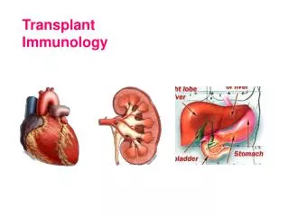
Transplant Immunology. Transplantation and Immunology. Review of cytokines Cells involved in alloreactivity Cell to cell interaction Major histocompatibility locus: transplant antigens Clinical immunosuppression. Sources and Effects of Cytokines. Cells involved in alloreactivity.

E N D
Transplantation and Immunology • Review of cytokines • Cells involved in alloreactivity • Cell to cell interaction • Major histocompatibility locus: transplant antigens • Clinical immunosuppression
Cells involved in alloreactivity • Key components • T cells • B cells • Antigen presenting cells (APC) • Development of lymphoid system begins with pluripotential stem cells in liver and bone marrow of fetus. As fetus matures, bone marrow is primary site for lymphopoiesis . • Pre T cells migrate to thymus, where CD3+ cells mature and become “educated” to self • Learn to restrict to self MHC and learn tolerance to self antigens • Mature T cells then populate lymph nodes, spleen, gut
Development of tolerance occurs centrally and peripherally • Central tolerance occurs through clonal deletion in the thymus • Pre-T cells (CD3+) enter thymus, proliferate and become CD4+ and CD8+ • Undergo “education” by self MHC class I or II
POSITIVE SELECTION • T cells that have a TCR receptor molecule with intermediate affinity for self MHC survive • If affinity too high or low, cells undergo apoptosis • NEGATIVE SELECTION • Occurs when T cells exposed to self antigens • Undergo apoptosis if react too strongly
Apoptosis • Regulated cell death • Cell condenses, fragments, phagocytosis occurs • Occurs through Fas Ligand system • Fas- receptor expressed on activated T cells • Expression of Fas and FasL lead to apoptosis
Cell to Cell Interactions • APCs- dendritic cells and macrophages- • Bind antigen and present it to B and T cells • Protein antigens need to be digested by phagocytes before presented to lymphocytes for self and non self recognition by MHC
T cell activation • TCR- T cell receptor recognizes antigens only if presented as peptide/MHC complexes presented on surface of APCs • TCR (heterodimer) binds covalently with CD3 molecule • Foreign antigen causes conformational change; causes intracellular signaling • When T cell activated, TCRs decrease; IL-2/IL-2R release increases(thru phospholipase C activation) • T cell proliferates
T cell activation also requires co-stimulatory signals (CD 28/B-7 interaction)Without this, T cell anergy results (basis for monoclonal Ab)
T cell effector functions • Cytotoxic T cells (CD8)- interact with MHC class I/peptide complexes and lead to cell lysis • Helper T cells (CD4)- recognize antigen in context of MHC class II molecules • Leads to cell mediated (TH1) or humoral (TH2) response
B lymphocytes • IL-7- growth factor for pre B cells • IL-4, 5, 6 stimulate maturation and proliferation of B cells • Responsible for antibody mediated immune response against foreign antigen • Express immunoglobulin antibody on cell surface • One antigen specific antibody produced per mature B cell • Naïve B cells express IgD and IgM • After antigen stimulation, undergo isotype switching to produce IgG (memory B cells)
Macrophages • Role of monocyte/macrophage • Phagocytosis • Presentation of processed antigen to lymphocytes results in production of cytokines
MHC locus/transplant antigens • Major histocompatibility locus located on Chromosome 6 • Produces Human Leukocyte Antigens (HLA) • Class I molecules are expressions of HLA-A, HLA-B, HLA-C • Class II molecules are expressions of HLA-DR, HLA-DQ, HLA-DP • Class III- contains mediators of immune function (TNF, heat shock protein)
Comparing MHC Class I and II Properties Class I Class II ANTIGENS HLA-A, B, C HLA-D-DR/Q/P TISSUE On all nucleated cells involved in DISTRIBUTION cells immune system FUNCTIONS Endogenous Ag Exogenous Ag presented to CD8 presented to (cytotoxic) T-cells CD4 (T-helpers)
HLA-Typing: Prevention and Rejection • Must make graft less antigenic so host doesn’t reject it • Major strategy: minimize alloantigen differences between donor and host • ABO compatibility to prevent hyperacute rejection • HLA (tissue) typing: HLA-A, HLA-B, HLA-DR most important • More alleles matched, greater survival of graft in 1st year • Appears to only matter if 6 allele match
HLA-typing • Serologic • Uses antigen specific serum to bind cells expressing antigen • Not very accurate • Molecular • PCR • Cross match • Uses flow cytometry to test for preformed antibodies
Rejection • Three types • Hyperacute- due to preformed Ab • ABO incompatibility • Minutes to hours • Prevented by screening • Result of prior pregnancy, transplant, blood transfusion • Cannot be treated with anti rejection meds • Mechanism- complement cascade; mediated by IgG • Characterized by rapid thromobosis/occlusion of graft vasculature
Acute rejection • Mediated by T lymphocytes • Occurs 1-3 weeks after transplantation without immunosuppression • Most common 3-6 months post transplantation but can occur anytime • Characterized by macrophage/lymphycyte infiltration
Acute Rejection • Two forms • Acute vascular rejection • IgG response • Leads to lysis of endothelial cells and cytokine production • Endothelial necrosis • Acute cellular rejection • Necrosis of parenchymal cells due to infiltration of T cells and macrophages
Chronic rejection • Appears as fibrosis and scarring • Atherosclerosis in heart patients • BOOP in lung patients • “vanishing bile duct syndrome” in liver patients • Fibrosis, glomerulonephropathy in kidney patients • Due to ischemia and inflammation
Chronic Rejection • Risk factors • Previous acute rejection ( risk with more episodes) • Inadequate immunosuppression • Initial delayed graft function • Donor factors (age, HTN) • Procurement issues (ischemia time, reperfusion injury) • Recipient issues (DM, HTN, infections)
Clinical immunosuppression • Risks of immunosuppression • Infection • Environmental pathogens and reactivation of host pathogens (CMV, EBV) • Requires prophylaxis with gangcyclovir; hep B vaccine, Bactrim to prevent PCP, UTI • Malignancy • No increase in lung, breast, prostate, colon, uterine CA • Skin tumors, cervical CA, Kaposi’s, lymphoma, other virus mediated tumors more common in Tx patients • Cardiovascular disease • Pretransplant risk factors (DM, HTN, CAD) amplified by immunosuppression • Pts need good pre-op workup
Objectives of Immunosuppression • Facilitate acceptance of the allograft • Specific • Low toxicity
Basic Strategies of Immunosuppression • Induction (High dose initial immunosuppression) • Facilitate graft acceptance • Minimize early rejection • Favor induction of tolerance • Maintenance therapy for chronic acceptance • Augmentation to reverse acute rejection.
Graft Rejection • Highly dependent on T cell activation and proliferation. • Signaling pathways and control points for entry into cell cycle are appropriate targets for immunsuppression.
Broad Mechanisms of Immunosuppression • T cell depletion. (induction) • Inhibition of T cell activation. • Block antigen binding. • Block accessory molecules. • Inhibition of IL-2 production. • Inhibition of T cell proliferation. • Inhibition of B cell proliferation.
Induction Agents-Thymoglobulin • Antithymocyte globulin- polyclonal sera produced when human lymphocytes injected into animals (rabbit or horse) • Interferes with cell-mediated reactions: allograft rejection, graft v host disease • Used in early post transplant period or to reverse ongoing rejection • Can cause anemia, thrombocytopenia • Allergic reaction most common problem
Induction Agents-Monoclonal Antibody • OKT3- engages CD3 TCR receptor • TCR receptor internalized by cell and no longer expressed on cell surface • T cells removed by spleen and reticuloendothelial cells • Less effective over time • Cytokine release syndrome
Induction Agents-IL-2R inhibitors • Basiliximab/ daclizumab (CD25 Ab) • Bind to IL-2 receptors without activating them • Cells can’t bind IL-2; can’t proliferate • Must be combined with other immunosuppressants • Well tolerated
Induction Agents- • Rituximab- Anti-CD 20 Ab • Treats humoral rejection due to B cell depleting ability • Campath 1H- Anti CD52 Ab • Acts against B/T cells, macrophages • Depletes lymphocytes for 2-6 months
Maintenance Agents • Adrenal Corticosteroid- prednisone • Antiproliferative • Azathioprine • Mycophenolate mofetil • Leflunomide • T cell directed • Calcineurin inhibitors: cyclosporine, tacrolimus • Cell cycle arrest: sirolimus • Lymphocyte Sequestration- FTY720
Adrenal Corticosteroids • Suppresses transcription and secretion of IL-1, IL-6, TNF • Inhibits IL-2 production and binding of IL-2R • Blocks ability of macrophages to respond to signals such as migration inhibition and activation factors • Side effects • Cushingoid features • Ulcer/GI bleed • Diabetes • Avascular necrosis
Antiproliferative AgentsAntimetabolites • Lymphocytes depend on de novo purine synthesis rather than salvage pathway • Azathioprine (6 mercaptopurine) • Blocks de novo purine synthesis, impedes T cell replication • Mycophenolate (Cellcept) • Blocks inosine monophosphate dehydrogenase (Rate limiting step for de novo sythesis) • Side effects- diarrhea, vomiting, bone marrow suppression, opportunitistic infections
Antiproliferative AgentsAntimetabolites • Leflunomide • Blocks dihydro-orotate dehydrogenase (necessary for de novo pyrimidine synthesis)
Inhibitors of T cell Receptor Signaling • Cyclosporine-binds to cyclophilin • Complex binds/inhibits calcineurin • Blocks cytokine production, esp IL-2, but does not inhibit proliferation • P 450 metabolism • Side Effects • Gingival Hyperplasia • Infection • Nephrotoxicity • Hypertension • Tremors, Nightmares, Insomnia • Hirsutism • Fibrous Breast Tissue
Inhibitors of T cell Receptor Signaling • Tacrolimus (FK506, Prograf) • Also inhibits calcineurin, thus inhibiting IL-2 production • Inhibits production of cytotoxic T cells • Side effects: alopecia, risk of post transplant diabetes
Inhibitors of T cell Receptor Signaling • Sirolimus • Macrolide antibiotic • Causes cell cycle arrest • Blocks transduction of signals from IL-2R to nucleus rather than blocking cytokine gene expression
T cell Signaling: Calcineurin Calcineurin
Lymphocyte Sequestration • FTY720 • Sequesters lymphocytes in Peyer’s patches • Can cause bradycardia