Download
1 / 20
250 likes | 451 Vues
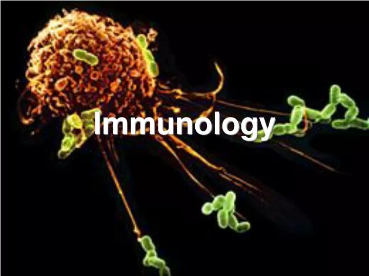
Immunology. Immunity – First Line of Defense (Keeping pathogens from getting in). Skin and mucous membranes Epithelium traps pathogen – swallow or expel them from the body Secrete oil and sweat – kills pathogens with a pH of 3-5

E N D
Immunity – First Line of Defense (Keeping pathogens from getting in) • Skin and mucous membranes • Epithelium traps pathogen – swallow or expel them from the body • Secrete oil and sweat – kills pathogens with a pH of 3-5 • Lysozyme is produced by eyes and upper respiratory tract – digests cell walls so kills bacteria
Non-Specific Cells (develop in bone marrow) Neutrophils Eosinophils Basophils Moncytes Natural Killer Cells Proteins Specific B-cells originate from bone marrow Make anti-bodies – destroy soluble pathogens T-cells Originate in bone marrow and then go to thymus T-killer – kill tumor cells and infected cells T-helper – activate T and B cells Immunity – InternalWhite Blood Cells/Proteins
Non-Specific Immunity • Quick, short-lived, activated specific immunity • Activated by damaged cells releasing chemical attractants or break in skin • Limits the spread of pathogens
Non-Specific Immune Cells • Neutrophils ( 60-70% of WBC’s) First responders • Enter tissue, phagocytize invaders, die in the process • Monocytes ( 5% of WBC’s) • Enter tissue, become macrophages (lg. phagocytes), kills cells by NO, reactive oxygen, and digestive enyzmes in their lysosomes • Releases cytokines to attract other immune cells • Later act as APC’s (antigen presenting cells) to specific immune cells
Non-Specific Cells continued • Eosinophils (1.5% of WBC’s) – release granules of enzymes to kill parasites • Natural Killer Cells – destroy infected cells and tumor cells by breaking open their cell membranes with a secreted enyzme • Basophils and Mast Cells – release histamine in response to physical damage or damage by microorganisms – Causes an inflammatory response
Inflammation • Histamine increases blood flow to an area by dilating the pre-capillary arteries and constricting the venules • Increases the permeability of bv so immune cells can get into the tissue • Blood leaks into the area causing redness and swelling • Clotting factors activate and complement attracts macrophages • Pus – dead WBC’s and fluid leaked from the bv. • Widespread inflammation – causes low blood pressure and high fever (some inflammatory cells release pyrogens that reset the brain’s thermostat) – can die of septic shock
Non-Specific Immunity - Proteins • Chemokines or Cytokines – released from damaged cells or other immune cells • Bind to receptors on immune cells • Cause neutrophils to be released from the bone marrow • Cause monocytes to produce NO • Cause basophils to produce histamine • Etc.
Proteins Continued • Complement – 20 proteins (both specific and non-specific) • Non-specific – attracts phagocytes, coats bacteria making it easier to phagocytize, binds to basophils and mast cells causing release of histamine and inflammation • Binds to 2 Ab bond to antigen and causes lysis of invaders • Interferon – produced by virus infected cells – makes nearby cells resistant to virus infection • Interleukins – activate other immune cells
Specific Immunity Uses Specific Antigens • Activity Immunity – your own immune system responds • Pasive Immunity – transfer of antibodies • Rabies Treatment • Mother’s Milk • Humoral – antibodies (proteins) – can only attack free antigens • Cell Mediated – can only attack cells infected with pathogens and cancer cells
General Information about Specific Cell Mediated Immunity • Primary response – takes 5-10 days for a maximum response against a specific antigen • Secondary response – takes 3-5 days for a maximum response (due to memory cells which divide faster and survive longer) • When a free antigen binds to a B cell – the B cell secretes soluble antibodies and undergoes mitosis • When an antigen on an infected cell binds to a T killer cell it makes clones of the T cell and secretes perforin to kill the infected cell • When an antigen on an APC binds to a T helper cell – The cell makes cytokines that cause specific T killer and B cells to multiply
MHC • MHC = major histocompatibility complex • Marker proteins on the cell’s surface for immune recognition • 200 MHC molecules • Class I MHC – on all cells • Class 2 MHC – on macrophages, B and T cells
T cells • T - Helper cells • CD4 cells (part of receptor) • Recognizes antigen and MHC class II (APC’s) • When binds to APC, makes cytokines that act on B and T cells (causes them to multiply and activate) • T – Killer cells (cytotoxic T cells) – Steps of Activation 1. Pathogen is eaten by a macrophage (Antigen Presenting Cell or APC) 2. APC digests the pathogen and displays antigens on MHC class II 3. T-helper binds and get activated – makes cytokines to activate T killer cells – only activates T killer as shown below 4. CD8 cells (part of receptor) • Recognizes infected or cancerous cells because they are displaying antigen on MHC class I (binds to the infected cells) • Releases enzymes that punch holes in the infected cell’s membrane • Cell explodes releasing pathogens • Pathogen no longer has a place to reproduce – antibodies pick it up or macrophages eat it Immunology You Tube Video
B cells • Has antibodies embedded in membrane as receptors / secretes Ab in soluble form when activated Steps of B cell activation • Pathogen is eaten by a macrophage (Antigen Presenting Cell or APC) • APC digests the pathogen and displays antigens on MHC class II • T-helper binds and get activated – makes cytokines to activate B cells • B cells bind to antigens on free pathogens that match the shape of their receptor (membrane antibodies) • Only the B cells that bind to antigen get activated by the cytokines – become plasma cells (make 2000 Ab/sec and memory cells (can multiply faster the second time)
Antibody Structure Variable Region makes different 3-D binding sites It is made up of three differerent regions: Variable region – 400 different choices Diverse region – 15 different choices Joining region – 4 different choices 1 trillion different shaped antibodies
B cells also become APC’s when activated and further activate T helpers which produce more cytokines to activate the B cells more
Antibody Functions • Bind to antigen (neutralization) – may block the activity (block virus from binding to a host cell, etc.) • Clumping or coating bacteria (opsonization) – makes it easier for macrophage to engulf • Activates complement which punches holes in the cell membrane of bacteria