Download
1 / 21
270 likes | 1.02k Vues
We Were Not Meant to Walk on Our Hands: The Gymnast’s Wrist. Lauren Grossman, MD Stony Brook Orthopaedic Associates: Division of Sports Medicine Sports Medicine Update: Women in Sports June 14, 2013 . Background. Many schools incorporate gymnastics

E N D
We Were Not Meant to Walk on Our Hands: The Gymnast’s Wrist Lauren Grossman, MD Stony Brook Orthopaedic Associates: Division of Sports Medicine Sports Medicine Update: Women in Sports June 14, 2013
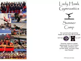
Background • Many schools incorporate gymnastics • About 600,000 students involved annually • Entry level around age 7 years of age • Peak involvement and performance around 13 years • All events place high levels of stress on the wrist joint • High level loading • Extremes of joint positioning
Background • Incidence of chronic injuries: 17-43% • 80-90%: chronic overuse at wrist • Injury rate in nationally ranked women’s collegiate gymnastics program: 0.95/participant/season • Level of competition , injury rates • Principle load type: compression • ***Dorsiflexion*** • Weight bearing joint • Degree, direction, and frequency of forces transmitted across the wrist • Superimposed on skeletal immaturity leads to problems
Chronic Osseous Injuries with Physeal Stress Reaction • Aka “Dorsiflexion Jam Syndrome” • Extreme dorsiflexion with axial compression and torsion on a chronic repetitive basis • Chronic stress adverse effect on endochondralossification • Repeated microtrauma disruption metaphyseal vascular network required for proper ossification • Transformation of cartilage to bone impeded widened irregular growth plate • Circulation reestablished, cartilage rapidly ossified • Usually no long term sequelae
Chronic Osseous Injuries with Physeal Stress Reaction • Presentation: • History: • Dorsal wrist pain • Swelling exacerbated by activities • Physical Exam: • Local swelling over dorsal distal radius • Exacerbation with axial loading and dorsiflexion
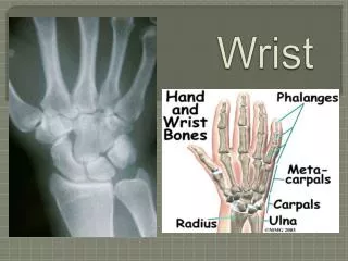
Chronic Osseous Injuries with Physeal Stress Reaction • Radiographic findings: • Widened distal radial physis • Cystic changes and irregularity of the metaphyseal margin • Palmar and radial beaking adjacent to the physis • Haziness of physis • Bone scintigraphy: increased uptake, often in both wrists
Chronic Osseous Injuries with Physeal Stress Reaction • Treatment: • Cornerstone = early diagnosis and cessation • No weightbearinguntil asymptomatic • FROM • No pain with provocative maneuvers (axial loading with forced dorsiflexion/handstand)
Consequences of Untreated Physeal Stress Reaction • Radiographic findings take longer to heal than if identified early • Radiographic healing around 3 months • No information if it must occur before return to full activity • Permanent alterations such as premature closure have been reported, leading to the ulnar plus variant
Ulnocarpal Impaction Syndrome • Presentation: • History: Ulnar sided wrist pain aggravated by activity • Physical Exam: • Tenderness over proximal lunate and triquetrum • Aggravated with ulnar deviation, especially with translation of ulnar head from anterior to posterior
Ulnocarpal Impaction Syndrome • Radiographic findings: • Positive ulnar variance • Rarefaction of triquetrum and lunate (impingement) • Diagnosis: arthrogram or arthroscopy can determine integrity of TFCC and triquetrolunateinterosseousmembrance
Ulnocarpal Impaction Syndrome • Treatment • Avoidance of aggravating activities • NSAIDS • Failure immobilization 4-6 weeks • If persistent: operative management • Arthroscopic debridement of TFCC tears, infrequently reported • Distal ulnar shortening or arthroscopic wafer resection for positive ulnar variance
Distal Radius and Ulna Physeal Injury from Distraction • Etiology: • Forces borne by digital flexors • Contraction causes a compression load to the radius and ulna • Dowel grips – decrease contraction forces required to maintain grip • Suspends the forearm and the body from the hand and the wrist • Traction induced distal radius physeal stress reaction • Differs from compression stress reaction - aggravated by the rings and high bar rather than pommel horse
Distal Radius and Ulna Physeal Injury from Distraction • Physical exam: provocation of symptoms with axial distraction • Treatment: • Cessation of aggravating activity • Avoidance of dowel grips • Evidence of complete healing after 4 months of treatment on x-ray
Scaphoid Impaction Syndrome • Etiology: • Repeated hyperextension of wrist impingement of dorsal rim of the radius and proximal carpal row • Pommel horse, floor exercises
Scaphoid Impaction Syndrome • Presentation: • History: pain with hyperextension at the wrist • Physical Exam: pain and point tenderness over dorsal rim of the scaphoid reproduced by hyperdorsiflexion • Radiographic findings: ossicles or hypertrophic dorsal scaphoid/radius rim on lateral
Scaphoid Impaction Syndrome • Treatment: • Symptoms always resolve with 2 weeks rest, temporary splinting and avoidance of hyperdorsiflexion stresses • Strengthening of the wrist and finger flexors with orthosis that limits dorsiflexion • Steroid injection may help ameliorate synovitis
Scaphoid Impaction Syndrome • Surgical exploration rarely necessary: may find chondromalacia, synovitis, capsular stripping, osteophytosis • Cheilotomy of either or both opposing surfaces • 2 weeks of SAC, graduated ROM program • No competitive level participation until FROM and strength compared to uninvolved side
Triquetrolunate Impaction Syndrome • Etiology: • Ulnardeviation and dorsiflexion • Floor exercises and pommel horse • Acute or chronic (racquet sports) injury
Triquetrolunate Impaction Syndrome • Presentation: • Physical exam: dorsoulnarswelling and point tenderness over triquetrolunate area • Reproduction of pain with ulnar deviation and dorsiflexion • Radiographic findings: • Dorsal triquetrum rim osteophytes/fractures
Triquetrolunate Impaction Syndrome • Treatment: • Rest, avoidance of aggravating activities • Surgical exploration: chondromalacia, synovitis, osteophytosis • Exposure of triquetrohamateand ulnocarpal articulations • Cheilectomy usually effective by removing 2-3mm of opposing rim surfaces • Synovectomy for inflamed tissues
Summary • Gymnastics participation common • Many injuries due to weightbearing through the wrist joint • Overuse can cause damage to physis • KEY: stop aggravating activities • Limited role for therapy • DO NOT resume activities until symptoms COMPLETELY resolve • Surgery last resort