Download
1 / 73
740 likes | 773 Vues
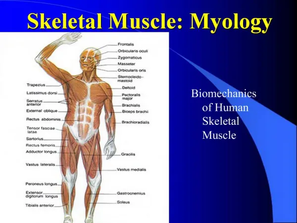
GENERAL MYOLOGY. MUSCULAR SYSTEM unit- muscle = musculus (myos) Active component of the locomotor system- it is controlled by nerves

E N D
MUSCULAR SYSTEM unit- muscle = musculus (myos) • Active component of the locomotor system- it is controlled by nerves • The main demonstration of mechanical function of muscle fibers (on the base of excitations comming through the motor nerve fibers)is their shortening– contraction (movement) • Contractile proteins myosin and actin, form the basis of myofibrils of muscle fibers
Heart muscle Smooth muscle Striated muscle
Function of the muscular system • motion function – muscular system constitues active component of the locomotor system • shape function - musculature forms exterior (external shape) of a man • termoregulation – it is releasing heat • It helps blood circulation • It keeps basic muscle tension ATTACHMENT To the bones: skeletal muscles- mm. sceleti- over 600 in the body, mostly paired, they form 1/3-1/2 of entire body weight To the skin: skin muscles- mm. cutanei- mainly on head and neck Relationship to organs: organ muscles To the articular capsules: mm. articulares
The internal structure of striated muscle 1) Striated muscle tissue (myosin and actin)- muscle fiber 2) Fibrous tissue(it covers the muscle fibers, primary and secondary fasciculi – important for metabolism between muscle fiber and blood circulation of muscle, on the surface, there is unbroken coveringfascia =fascia 3) Logisticcomponents (vessels and nerves) 4) Special apparatus
INTERNAL STRUCTURE OF MUSCLE Muscle part: the basic structural and functional unit of muscular system, it is the muscular fiber, which was created by fusion of many consecutive cells= multinucleated formation • The fibers inside muscle have following arrangement– form muscle fasciculi, they combine into bundles until they create the entire muscle • Muscle fibers are interconnected with thin collagenous fibrous tissue, so-called perimysium internum (endomysium) • the surface of whole muscle is covered by fibrous tissue, so-called perimysium externum (epimysium)
Tendon: tendon is created by regularly arranged fibers of tough collagenous fibrous tissue, which have hierarchical arrangement – single fibers combine to fasciculi, then to larger bundles until they form the whole tendon • fibers are connected with thin collagenous fibrous tissue, so-called peritenonium internum (endotenonium) • On the surface, there is a fibrous covering, so-called peritenonium externum (epitenonium) • aponeurosis- flat tendons
EXTERNAL STRUCTURE OF MUSCLE • origin (origo): part of the muscle that runs from bone (or skin); it is the place, where the muscle doesn´t change its position during contraction (so-called: fixed point- punctum fixum), it is usually formed by tendon • belly (venter): fleshy part of muscle, its beggining is called caput (head), its end is called cauda(tail) • insertion (insertio):is formed by tendon; it is the place, where the muscle changes its position during contraction (so-called: mobile point- punctum mobile), the tendon attaches usually to a bone, sometimes to skin or organ
CLASSIFICATION OF MUSCLES • ACCORDING TO PREVAILING SIZE • Long muscles:theyhaveribbon-likeorrope-liketendons • Shortmuscles: theyhaveribbon-likeorrope-liketendons • Flatmuscles:theyusuallyhavewideflattendons= aponeurosis • Roundmuscles: ring-likeshape, theyencirclesomeopenings, they are narrowingduringcontraction
3. ACCORDING TO A NUMBER OF HEADS • Muscles with one head: one head • Muscles with more heads: more heads (more origins), which connect into one muscle belly. (musculus biceps, musculus triceps,musculus quadriceps) 4. ACCORDING TO A NUMBER OF BELLIES • With one belly: only one belly • With more bellies: two or more consecutive bellies, which are separated from each other by tendons (tendo intermedius)
CLASSIFICATION ACCORDING TO FUNCTION • Muscle can make its function, only if it span minimal one movable bone junction • muscle making specific movement is calledagonist (executor) • Muscle which participate in some movement are called synergists • Muscles making opposite movement are called antagonists flexors× extensors adductors× abductors sphincters× dilatators pronators× supinators levators× depressors erectors elevators tensors
Contraction Isotonic: change of lenght concentric: shortens excentric: extends Izometric: change of tension
Vessels-blood and lymphatic- nutritionofmuscle. They enter themuscle in place called porta musculi (hilus musculi)- neurovascular hilus. Nerves - diploneuralmuscles- innervatedfromtwonerves • plurineuralmuscles- innervatedfrom more nerves -motor fibers:theybringimpulsesforcontractionofmusclefibers, they are terminated as motor plates on themusclefiber - Senzor fibers:bringinformationfrommuscleintocentralnervoussytem, aboutpain, tension.
SPECIAL APPARATUS 1. Fascia (fasciae): fibrous membranes, which cover one whole muscle or group of some muscles. Septa intermuscularia- separates single groups of muscles, they are attached to a bone Retinacula- eyelets, which holds muscle tendons to a bone. 2. Synovial bursae(bursae synoviales): pouches around the joint, derivatives of the joint capsule, in the places, where tendons and muscle lie directly on the bone 3. Synovial sheath (vaginae tendinum): cover long tendons of muscles in areas exposed to mechanical loading. Layer- superficial- vagina fibrosa- peritenonium - deep- vagina synovialis- epitenonium
M. temporalis origin: linea temporalis inferior, temporal fascia insertion: processus coronoideus mandibulae innervation: N. trigeminus (nn. temporales profundi from 3rd branch) function: elevation, partly retraction of mandible
origin: arcus zygomaticus and os zygomaticum insertion: tuberositas masseterica innervation: N. trigeminus (n. massetericus from 3rd branch) function: elevation of mandible, chewing movements
3) M. pterygoideus medialis origin: fossa pterygoidea and tuber maxillae insertion: tuberositas pterygoidea innervation: N. trigeminus (n. pterygoideus medialis from the 3rd branch) function: elevation of mandible 4) M. pterygoideus lateralis origin: lamina lateralis processus pterygoidei, facies infratempotalis alae majoris ossis sphenoidalis insertion: fovea pterygoidea mandibulae innervation: N. trigeminus (n. pterygoideus lateralis from the 3rd branch) function: by double-sided contraction: protraction of mandible
Mimic muscles m. occipitofrontalis m. temporoparietalis Muscles of palpebral fissure m. orbicularis oculi m. depressor supercilii m. corrugator supercilii m. procerus
3) Muscles of the mouth m. orbicularis oris m. depressor anguli oris m. depressor labii inferioris m. risorius m. levator labii superioris alaeque nasi m. levator labii superioris m. zygomaticus major m. zygomaticus minor m. levator anguli oris m. buccinator m. mentalis 4) Muscles of the nose m. nasalis m. levator labii superioris alaeque nasi
Head fasciae Fascia temporalis – together with skull bones, it creates a cavity for m. temporalis Fascia masseterica – continues as fascia parotideomasseterica (to the gland) Fascia buccopharyngea – from the lips to pharynx
Musculi colli (muscles of the neck)
Platysma • Subcutaneous muscle, on superficial cervical fasciafrom clavicle to the face O: fascia pectorialis, fascia deltoidea I: skin of the face F: it stretches cervical skin IN: ramus colli n. facialis
M. sternocleidomastoideus O: manubrium sterni, sternal end of calvicle I: processus mastoideus, external edge of linea nuchae superior F: at unilateral contraction – lateroflexion, bilateral contraction – retroflexion, auxiliary inspiratory muscles (at fixed head and cefvical spine) IN: n. accessorius + C2 - C4
Musculi suprahyoidei M. DIGASTRICUS M. STYLOHYOIDEUS M. MYLOHYOIDEUS M. GENIOHYOIDEUS
M. DIGASTRICUS Muscle with two bellies O:venter anterior: fossa digastrica, it is changing into tendon on hyoid bone, continues as venter posterior I: incisura mastoidea F: depression of mandible, elevation Of hyoid bone I: venter anterior - n. mylohyoideus (n. trigeminus) venter posterior - n. facialis
M. STYLOHYOIDEUS Through its cleft tendon m. digastricus passes O: processus styloideus I: body of the hyoid bone F: it elevates the hyoid bone during swallowing I: n. facialis
M. MYLOHYOIDEUS Forms the flexible bottom of the mouth- diphragma oris O: linea mylohyoidea I: os hyoideumraphe mylohyoidea - fibrous connection of both muscles F: depression of mandible at fixed mandible, elevation of hyoid bone I: n. mylohyoideus (n. trigeminus)
M. GENIOHYOIDEUS Above m. mylohyoideus O: spina mentalis I: body of the hyoid bone F: it participates in forming of the bottom of the mouth I: fibers from C1
Mm. infrahyoidei 1. m. sternohyoideus 2. m. sternothyroideus 3. m. thyrohyoideus 4. m. omohyoideus F: they fix the hyoid bone, they participate in swallowing reflex I: ansa cervicalis profunda C1 - C3 - except m. thyrohyoideus -> C1
M. STERNOHYOIDEUS O: dorsal surface of manubrium sterni + sternalnal end of clavicle Ú: body of hyoid bone
M. STERNOTHYROIDEUS behind m. sternohyoideus and more laterally O: manubrium sterni and 1st rib I: linea obliqua
M. THYROHYOIDEUS O: linea obliqua on cartilago thyroidea I: cornu majus of hyoid bone
M. OMOHYOIDEUS With two bellies O: venter inferior- margo scapulae sup., bellow m. sternocleidomastoideus it continues as a tendon and then int chnages into venter superior I: body of hyoid bone