Download
1 / 40
410 likes | 606 Vues
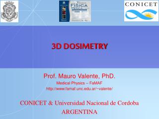
Energy dependence of detectors for 3D dosimetry of light ion beams. Hugo Palmans MedAustron , Wiener Neustadt, Austria and National Physical Laboratory , Teddington, UK. Overview. Need for measuring absorbed dose Characteristics of ideal 2D and 3D detectors

E N D
Energy dependence of detectors for 3D dosimetry of light ion beams • Hugo Palmans • MedAustron, Wiener Neustadt, Austria • and National Physical Laboratory, Teddington, UK
Overview • Need for measuring absorbed dose • Characteristics of ideal 2D and 3D detectors • Energy dependence of detector response to absorbed dose
Absorbed dose versus fluence • Most of this conference:, or • This presentation: • energy imparted • thermalisation • ionisation • chemical states • physical states
probability of tumour control 1.0 probability of severe complications (x-rays) 0.8 probability of tumour control without severe complications (x-rays) probability of severe complications 0.6 (protons) Probability probability of killing tumour without severe complications (protons) 0.4 0.2 0.0 0.0 20.0 40.0 60.0 80.0 100.0 Dose (Gy = J kg-1) Need for accurate dosimetry
Need for measuring absorbed dose • At the cellular level: • direct DNA damage: ionisation at nanoscale • indirect DNA damage: ionisation at microscale • other damage: tens of micrometers scale • bystander effects: millimetre scale • For photons and electrons: • biological effects ~ ionisation ~ absorbed dose • For protons and ions: • biological effects ~ ionisation * wi ~ absorbed dose * wi * wD
Need for 3D dosimetry • Homogeneity • Target coverage • Cold spots • Out-of-field dose • Integral dose
Requirements • High spatial resolution • Small dosimetric “voxels” • Ease of operation, non-toxic • Reasonable cost • Fast readout • Stable in time; reproducible • Signal proportional to dose (or known functional relation) • Dose rate independent, large dynamic range • Orientation independent • Water-equivalence • Minimal/managable perturbing
passive (scattered) – dynamic (scanned) range modulator
ΔT/D a c (J·kg-1·K-1) (mK·Gy-1) (m2·s-1) water 4180 0.24 1.44×10⁻⁷ graphite 710 1.41 0.80×10⁻⁴ Calorimetry (thermalisation) Dmed=cmed·ΔT
0.6 mm thermistor 0.5 mm Calorimeters - water vs graphite
protons (100 MeV) 60Co (1MeV) 3,0 H+ OH• 2,0 OH¯ e¯aq G (100 eV-1) H2 H2O2 1,0 HO2 H• 0,0 10-1 100 101 102 LET (keV.mm-1) Water calorimeters – energy independent chemical heat defect? Ross et al (1996) Phys. Med. Biol. 41:1 Brede et al (2006) Phys. Med. Biol. 51:3667
Graphite calorimeters – energy independent dose conversion? ?
= D D s p w air w, air Q W Q W = = D air r m e V e air air Dose determination with ion chamber • Q: charge produced in the air of the chamber • W: mean energy required to produce an ion-pair in air Unfortunately, for commercially available chambers, the volume V is not known with the necessary accuracy (would otherwise be a primary standard!). We have to rely on methods other than “first principles”, which involve the use of ion chamber calibration factors
r = 1mm r = 1mm 1.30 o,p o,e 1.20 s 1.10 w,air 1.00 sw,air protons sw,air electrons 0.90 Water/air stopping power ratio ICRU 49 ICRU 37 103 ) -1 g 102 2 (MeV cm r / proton water 101 coll protons air S electrons water electrons air 100 10-6 10-4 10-2 100 102 Medinet al. 1997, Phys Med Biol 42:89 t = E /E k rest
p x 70.0 (Gy) water 60.0 2 Rsleeve sleeve 50.0 Rwall wall 40.0 Rcav air per proton per cm 30.0 c.e. Rcel z 20.0 Proton air 10.0 D 0.0 5.0 reconstruction Difference pdd and 0.0 -5.0 0.0 10.0 20.0 30.0 z0 Depth (mm) Perturbation correction factors Palmans 2011 Proc IDOS IAEA-CN182-230 Palmans 2006 Phys Med Biol 51:3483
Ionisation chambers – overall conversion air to dose Palmans, Dosimetry, in : Proton Therapy Physics, Ed Paganetti
Ionisation chambers (& any ionisation detector) – charge recombination • Palmans et al 2006 NPL report DQL-RD003
Initial recombination • Volume recombination (pulsed) (continuous) • Charge multiplication Ion chambers - recombination HT CE E
diamond detectors Fidanzioet al 2002 Med Phys29:669
Silicon-based detectors Kohno et al 2006 Phys Med Biol51:6077
TLD - protons Bessereret al 2001 Phys Med Biol 46:473
NPL therapy level alanine/EPR • Operates since 1991 • Bruker ESP 300 X-band 9” magnet • Pellets • 90% alanine + 10% paraffin wax • 5 mm diameter • 0.5 mm and 2.5 mm nominal thickness • Measurement reproducibility of • 2.5 mm pellets ~ 0.05 Gy
Alanine/EPR dosimetry Bassler et al, NIMB 266 929-936, 2008 CERN anti-proton beam Birmingham 15 MeV beam GSI 12C ion beam Herrmann et al 2011 Med Phys 38:1859
Radiochromic film Piermatteiet al 2000 Med Phys27:1655
Radiochromic film – energy dependence • Kirby et al. 2010 Phys Med Biol 55:417
An interesting one… depth dose distribution for fluencedetermination • Pic laser induced beam • Kirby et al. 2011 Laser Part Beams 29:231
Polymer gel dosimetry • Palmans et al 2006 NPL report DQL-RD003
Polymer gel dosimetry • BANG3-Pro2: • Zeidan et al. 2010 Med Phys 37:2145
Plastic scintillators • Liu et al. 2011 Phys Med Biol 56:5805 A. Beierholm, Risoe • Goulet et al. 2012 Med Phys 39:4840
Scintillators Safaiet al. 2004 Phys. Med. Biol. 49:4637
Microdosimetry… Superconducting Absorber Chip : about 5x5 mm2 TE Absorber SQUID Junctions Seb Galer, NPL Andrea Pola, Politecnico di Milano
Nanodosimetry… • Ion counter / PTB Startrack / INFN
Reading • C. P. Karger, O. Jäkel, H. Palmans and T. Kanai, “Dosimetry for Ion Beam Radiotherapy,” Phys. Med. Biol. 55(21) R193-R234, 2010 • H. Palmans, A. Kacperek and O. Jäkel, “Hadron dosimetry” In: Clinical Dosimetry Measurements in Radiotherapy (AAPM 2009 Summer School), Ed. D. W. O. Rogers and J. Cygler, (Madison WI, USA: Medical Physics Publishing), 2009, pp. 669-722 • H. Palmans, “Dosimetry,” In: Proton Therapy Physics, Ed. H. Paganetti (London: Taylor & Francis), 2011, pp. 191-219 • H. Palmans, “Monte Carlo for proton and ion beam dosimetry,” In: Monte Carlo Applications in Radiation Therapy, Ed. F. Verhaegen and J Seco, (London: Taylor & Francis), 2013, pp. 185-199