Download
1 / 1
50 likes | 356 Vues
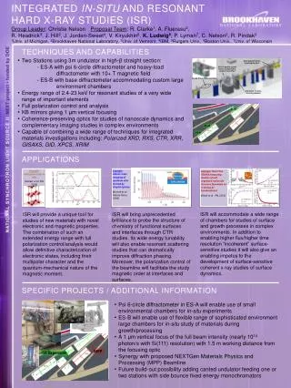
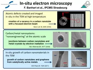
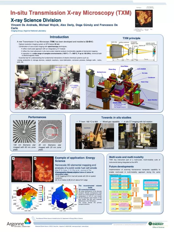
60 nm. 30 nm. In-situ Transmission X-ray Microscopy (TXM). X-ray Science Division. Vincent De Andrade, Michael Wojcik , Alex Deriy , Doga Gürsŏy and Francesco De Carlo Imaging Group , Argonne National Laboratory. Introduction

E N D
60 nm 30 nm In-situ Transmission X-ray Microscopy (TXM) X-ray Science Division • Vincent De Andrade, Michael Wojcik, Alex Deriy, DogaGürsŏy and Francesco De Carlo Imaging Group, Argonne National Laboratory Introduction • A new Transmission X-ray Microscope (TXM) has been developed and installed at 32-ID-C: • Highest resolution imaging system at APS (below 20 nm) • Combination of nano-scale imaging with spectroscopytechniques: • offers multi-scale approach with an integrated µ-CT module • offers the most advanced in situnano-scale material analysis functionality capable of elemental mapping • operation in a wide range of samples environments (-160°C < T < 900°C, P up to 100 GPa), chemical bath & electromagnetic fields • Essential tool for understanding the fundamental interactions of electro-chemical systems such as: • energy production & storage devices, catalytic reactions, nano-fabrication, corrosion process, biologic cells , rocks, soils, etc. TXM principle Performances Towards in situstudies • Mixed gas capability • in situ CT imaging with full rotation range • Tofrom -160 °C to 900 °C cryostream infrared heater X-rays X-rays 100 nm Siemens star imaged with 60 nm zone plate. 30 nm Siemens star imaged with 20 nm zone plate. Former 32-ID TXM Former 32-ID TXM Multi scale and multi modality TXM: key instrument part of a multi-scale, multi-modality suite of instruments being integrated at the APS • Future developments • Implementation of scanning fluorescence nanoprobe capability to enable multi-scale & multi-modality approach during the same experimental run • Example of application: Energy Science • Nanoscale 3D elemental mapping and analysis of a solid oxide fuel cell anode • After K. N. Grew, Y. S. Chu et al., Journal of The Electrochemical Society, 2010. B A 2.5 µm 2 tomographic dataset acquired above & below Ni absorption edge: 3D mapping of Ni in fuel cell anode with 38 nm spatial resolution [A] 16 eV below & [B] 24 eV above Ni K-edge The reconstructed volume feeds3D numerical models: 3D scalar distributions of the discrete transport. Results correspond to total fluxes at a current density of 3 A/cm2, temperature of 1073 K, and include the [C] hydrogen mole fraction in the pore phase and [D] ionic potential differences in the YSZ phase. The arrows denote the primary transport direction. C D The Advanced Photon Source is funded by the U.S. Department of Energy Office of Science Advanced Photon Source • 9700 S. Cass Ave. • Argonne, IL 60439 USA • www.aps.anl.gov • www.anl.gov