Download
1 / 43
430 likes | 449 Vues
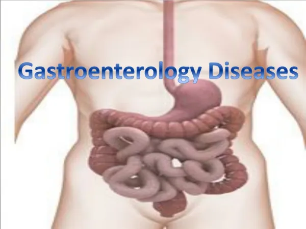
Gastroenterology 2. Diagnostic methods for gastrointestinal diseases. Laboratory investigations. ESR: increased: inflammation, tumors (but can be normal) Blood count leukocytes: : inflammation eosinophilia: helminthiasis, allergy

E N D
Laboratory investigations • ESR: increased: inflammation, tumors (but can be normal) • Blood count • leukocytes: : inflammation • eosinophilia: helminthiasis, allergy • anemia (Hb, HCT): GI bleeding (manifest or occult) • Se Iron : bleeding, malabsorption, chr.infection
Laboratory investigations • Liver tests: • AST(GOT), ALT(GPT): cell damage • ALP, GGT, bilirubin: cholestasis • prothrombin time, se albumin : liver failure • Pancreas: amylase, lipase, functional tests • Fecal occult blood test (FOBT) • Stool cultures for bacteria and parazites • Urine: jaundice, uroinfection, kidney stone • Duodenal aspiration
Abdominal ultrasound • features • specific US methods • Doppler-ultrasound - for vascular lesions • US-guided biopsy • EUS- endoscopic ultrasound - endosonography
Abdominal ultrasound • Liver • echogenity, masses, cysts, bile ducts, veins • Biliary tract • gallstones (hyperechoic lesion with acoustic shadow), sludge, CBD stones, cholecystitis • Pancreas • acute pancreatitis, chr.pancreatitis, pseudocysts, tumors • Others • ascites, organomegalies, lymph nodes, appendicitis, intraabdominal masses (tumor, abscess, cyst, inflammatory mass), kidneys
Radiology • Plain abdominal X-ray • free air (upright position) • gas/fluid levels within dilated loops • calcifications • Upper GI barium radiography (single or double contrast studies) • esophagus (first examination in dysphagia) • contour, peristalsis, folds • motility disorders, stenoses
Radiology • Upper GI barium radiography • stomach and duodenum • peristalsis, emptying, shape, folds, retrogastric space • perforation: with water-soluble contrast agent • in case of GI hemorrhage: endoscopy • Barium study of the small bowel • small bowel follow through study • enteroclysis • stenoses, polyps, mucosal alterations, ileitis terminalis
Radiology • Barium enema (double-contrast) (synonims: irrigoscopy, colonography) • mostly in cases of stenosis on endoscopy
Radiology - angiography • diagnosis of vascular diseases, obscure GI bleedings • therapeutic angiography is evolving (chemoembolisation of tumors, occluding bleeding vessels, dilation of vessels)
Computer tomography • features • specific CT methods • spiral/helical CT • contrast agents (orally administered, iv.) • CT-guided biopsy • virtual colonoscopy
Computer tomography • Liver • masses (benign, malignant [primary or metastatic neoplasms], hemangiomas, cysts, abscesses) , cirrhosis, ascites and other signs of portal hypertension, lymph nodes • Biliary tract • dilated bile ducts, imaging of CBD, distal bile duct stones, CBD neoplasms
Computer tomography • Pancreas - (the most useful method) • neoplasms: diagnosis, staging • acute pancreatitis: extent of necrosis, peripancreatic fluid collections, guided biopsies • chr. pancreatitis: pseudocysts, calcifications • Miscellaneous • staging of gastrointestinal malignancies, intra-abdominal masses (abscess, inflammatory, tumors), invasion of adjacent structures
Magnetic resonance imaging • generally not superior to CT in abdominal diseases • sensitive • very expensive • special methods • MR angiography • MRCP - magnetic resonance cholangio-pancreatography
Endoscopy • features • diagnostic endoscopy • provides histological sampling (biopsy, brush cytology) • therapeutic endoscopy
Upper GI endoscopyEsophagogastroduodenoscopy (EGD) • Diagnostic • GI bleeding • refractory vomiting • dysphagia, odynophagia • gastroesophageal reflux • ulcers • suspicion of neoplasm (weight loss, etc.) • surveillance of healing lesions • surveillance of polyps, tumors
Upper GI endoscopy • Therapeutic • treatment of variceal and nonvariceal GI bleeding • injection technics, hemoclip, ligation, thermal technics (elelctrocoagulation, heat probe, laser, argon plasma) • removal of polyps, early neoplasms • dilation of strictures • placement of feeding gastrostomy tube • removal of foreign bodies
Lower GI endoscopyColonoscopy, rectosigmoidoscopy, rectoscopy • Diagnostic • Bleedings (occult or hematochezia, iron deficiency) • Chronic diarrhea • Suspicion of cancer • Suspicion of inflammatory bowel disease • Screening for cancer (altered bowel habits, risk groups for colon cancer)
Lower GI endoscopyColonoscopy, rectosigmoidoscopy, rectoscopy • Therapeutic • Removal of polyps, early cancers • Dilation of stenoses • Decompression
Endoscopic retrograde cholangio-pancreatography - ERCP • Diagnostic • suspicion of choledocholithiasis • unexplained jaundice and cholestasis • acute gallstone pancreatitis • some cases of chr. pancreatitis • Therapeutic • endoscopic sphincterotomy - EST • endoscopic biliary/pancreatic drainage • endoscopic biliary/pancreatic stenting • dilation of strictures • endoscopic lithotripsy
Miscellaneous diagnostic methods • Biopsies (US/CT-guided)- liver, pancreas, masses • Punctions - ascites, cysts • Percutaneous transhepatic cholangiography (PTC) or drainage (PTD) • Laparoscopy • Helicobacter pylori diagnostics • stains, rapid urease-test, urease breath test (UBT) • 24h pH monitoring • Manometry(esophageal, rectal, Oddi-sphincter, bowel)
Gastroesophageal reflux disease - GERD • History: • Esophageal:heartburn, chest pain, regurgitation, acidic taste in mouth, dysphagia, odynophagia, Extraesophageal: chr.cough, asthma, noncardiac chest pain • Characteristics: increase in laying position night symptoms resolve after antacids • Physical findings: • Diagnosis: history, endoscopy, pH-monitoring, barium swallow
Esophageal cancer • History: dysphagia, odynophagia, pain, vomiting, weight loss • Characteristics: older males, alcoholics, smokers progressive dysphagia (solidsofterliquid) vomiting just after meals • Physical finding: general tumor signs • Diagnosis: barium swallow, endoscopy
Peptic ulcer (duodenal, gastric) • History: epigastric pain • Characteristics: • radiates to the back • duodenal: younger people, hyperacid symptoms, relapsing disease, more symptoms in spring and fall, pain resolves after meals and recur after 2 hours, night pain, resolve using antacids • gastric: older people, pain just after meals, weight loss • smokers • NSAID (aspirin) use
Peptic ulcer (duodenal, gastric) • Physical finding: epigastric/RUQ tenderness • Diagnosis: endoscopy
Peptic ulcer - complications • Bleeding:melena, hematemesis, (rarely: hematochezia) rectal digital examination • Perforation:acute onset very sharp pain (knife-like) liver/splenic dullnes: absent peritoneal signs: defence (guarding), rebound tenderness, no bowel sounds Dg: abdominal plain film study with water-soluble contrast agent
Peptic ulcer - complications • Obstructiona. reversible b. irreversible (scar) History: vomiting of undigested food fullness, pain Physical signs: succussion splash tenderness Diagnosis: gastric emptying study (barium) endoscopy
Gastric cancer • History:epigastric pain, fullness, vomiting, weight loss • Characteristics:older people, pain arise at meals dull, progressive pain • Physical findings:epigastric pain, epigastric mass Virchow’s lymph node general tumor signs occult bleeding • Diagnosis: endoscopy, US
Acute appendicitis • History: first periumbilical, later ileocecal pain nausea subfebrility • Characteristics: invariable first colicky, than steady pain • Physical findings: ileocecal tenderness (McBurney’s point) ileocecal guarding rebound tenderness obturator sign: pain rotating the right hip psoas sign: pain raising against resistance the straightened right leg • Diagnosis: physical examination, US, laboratory
Intestinal obstruction (ileus) 1. Mechanical • History: altered bowel habits, constipation, fullness, meteorism, cramping pain, vomiting (bile, fecal material) • Characteristics: variable or progressive • Physical finding: meteorism increased bowel sound splash signs of underlying disease • Diagnosis: plain abdominal x-ray, CT searching for the cause
Intestinal obstruction (ileus) 2. Paralytic • History:signs of the underlying disease, constipation, fullness, meteorism, cramping pain, vomiting • Physical finding:meteorism absent bowel sound splash signs of the underlying disease • Diagnosis: plain abdominal x-ray searching for the cause
Colorectal cancer • History: altered bowel habits bleeding (occult or manifest) late: signs of obstruction cramping pain general tumor signs positive family history • Physical finding:rectal digital examination late: mass, ileus • Diagnosis:endoscopy, US, CT
Acute hepatitis • History: asymptomatic after flu-like symptoms jaundice anorexia, dyspepsia RUQ pain • Physical finding: jaundice enlarged liver: smooth, soft, round, tender • Diagnosis: liver tests, virus tests
Chronic hepatitis • History: symptoms: not characteristic anorexia, dyspepsia later: symptoms of cirrhosis • Physical finding: enlarged liver (can be normal) • Diagnosis: US, liver biopsy, serology
Liver cirrhosis • History: alcohol consumption, chr. hepatitis (HBV, HCV, HDV, HGV, autoimmune), anorexia, dyspepsia, nausea ascites, edemas, portal encephalopathy jaundice, bleeding • Physical findings: first: enlarged liver micronodular: alcoholic macronodular: chr. virus or autoimmune hepatitis- postnecrotic cirrhosis end stage: small liver
Liver cirrhosis • Physical findings: skin: palmar and plantar erythema spider naevi icterus (scratching) gynecomasty testicular atrophy signs of portal hypertension: ascites (transsudate) caput Medusae splenomegaly edema • Diagnosis: US, liver biopsy, laboratory
Biliary colic • History:pain after fatty meals nausea, vomiting (often bile) fullness, meteorism • Characteristics: RUQ-pain, radiates to the back (scapula, right shoulder) mostly females • Physical finding: RUQ tenderness • Diagnosis: US
Acute cholecystitis • History: like in biliary colic + fever • Physical finding: Murphy’s sign • Diagnosis: US, laboratory: signs of inflammation Choledocholithiasis • History: like in biliary colic + obstr. jaundice • Diagnosis: US, ERCP, CT
Acute pancreatitis • History: gallstone disease,fatty meal,alcohol epigastric pain fullness, nausea, vomitus fever jaundice hypotony, shock • Characteristics: band-like, cramping pain radiates to the back
Acute pancreatitis • Physical findings: epigastric tenderness/guarding peritoneal signs signs of paralytic ileus (meteorism, no bowel sounds) skin signs: Cullen’s sign-periumbilical ecchymoses Grey-Turner’s sign- lumbar ecchymoses • Diagnosis: pancreatic enzimes, US, CT
Chronic pancreatitis • History: cramping pain anorexia, dyspepsia, nausea, vomitus gallstone or alcohol consumption weight loss steatorrhea • Characteristics: pain in the back increases after meals • Physical finding: epigastric tenderness epigastric mass (pseudocyst) sometimes jaundice • Diagnosis: plain abd. X-ray, US, CT, ERCP