Download
1 / 110
1.11k likes | 1.32k Vues
3. Gastroenterology. Learning Objectives. Identify the structures of the gastrointestinal system. Describe the process of digestion. Describe common gastrointestinal diseases and conditions, laboratory and diagnostic procedures, medical and surgical procedures, and drug categories.

E N D
3 Gastroenterology
Learning Objectives • Identify the structures of the gastrointestinal system. • Describe the process of digestion. • Describe common gastrointestinal diseases and conditions, laboratory and diagnostic procedures, medical and surgical procedures, and drug categories.
Learning Objectives • Give the medical meaning of word parts related to the gastrointestinal system. • Build gastrointestinal words from word parts and divide and define gastrointestinal words. • Spell and pronounce gastrointestinal words.
Learning Objectives • Analyze the medical content and meaning of a gastroenterology report. • Dive deeper into gastroenterology by reviewing the activities at the end of this chapter and online at Medical Terminology Interactive.
Multimedia Directory Slide 10 Digestive System Animation Slide 45 Digestive System Animation Slide 51 GERD Video Slide 57 Appendicitis Animation Slide 91 Ultrasound Video Slide 93 MRI Video Slide 103 Sigmoidscopy Video Slide 110 Medical Assisting Video
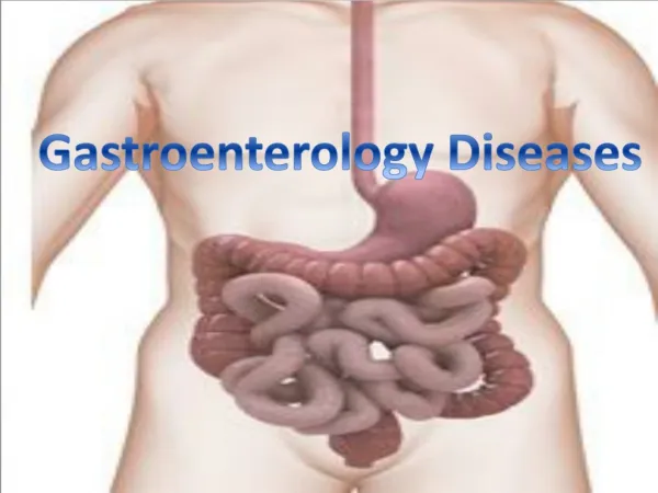
Gastroenterology The medical specialty that studies the anatomy and physiology of the gastrointestinal system and uses diagnostic tests, medical and surgical procedures, and drugs to treat gastrointestinal diseases.
Anatomy and Physiology Gastrointestinal System Begins at the mouth, continues through the thoracic cavity, and fills most of the abdominal cavity Upper gastrointestinal system includes the structures from the mouth through the stomach
Anatomy and Physiology (con’t) Gastrointestinal System (con’t) Lower gastrointestinal system includes the small and large intestines Purpose is to digest food, absorb nutrients, and remove undigested material (waste) from the body
Digestive System Animation Click on the screenshot to view an animation showing a tour of the digestive system. The animation may take a few seconds to start playing. Back to Directory
Anatomy of the Gastrointestinal System Oral Cavity and Pharynx Begins in the mouth, or oral cavity Oral cavity contains the teeth; tongue; hard palate; and soft palate with its fleshy, hanging uvula. Receptors on the tongue perceive taste and send this information to the gustatory cortex in the brain.
Anatomy of the Gastrointestinal System (con't) Oral Cavity and Pharynx (con’t) Lined with mucosa, a mucous membrane that produces thin mucus. The sight, smell, and taste of food cause the salivary glands to release saliva into the mouth; this moistens foods as they are chewed and swallowed. Saliva also contains an enzyme that begins the process of digestion.
Anatomy of the Gastrointestinal System (con't) Oral Cavity and Pharynx (con’t) There are three pairs of salivary glands: the parotid glands, the sublingual glands, and the submandibular glands. The teeth tear, chew, and grind the food during the process of mastication. The tongue moves food toward the teeth and mixes food with saliva.
Anatomy of the Gastrointestinal System (con't) Oral Cavity and Pharynx (con’t) Swallowing or deglutition moves food into the throat or pharynx. When food is swallowed, the epiglottis closes the entrance to the larynx, so that food in the back of the throat, pressing on the uvula, does not initiate the gag reflex.
Anatomy of the Gastrointestinal System (con't) Esophagus A flexible, muscular tube that connects the pharynx to the stomach. Lined with mucosa that produces mucus. By coordinated contractions of its wall—the process of peristalsis—food moves toward the stomach.
Anatomy of the Gastrointestinal System (con't) Stomach A large, elongated sac in the upper abdominal cavity that receives food from the esophagus. Divided into four areas: the cardia, fundus, body, and pylorus. The gastric mucosa is arranged in thick, deep folds known as rugae which expand as the stomach fills with food.
Anatomy of the Gastrointestinal System (con't) Stomach (con’t) The mucosa produces mucus that protects the lining of the stomach from the acid the stomach produces. Two sphincters (muscular rings) keep food in the stomach. The lower esophageal sphincter is located in the distal esophagus.
Anatomy of the Gastrointestinal System (con't) Stomach (con’t) The pyloric sphincter is located in the distal end of the stomach. Chyme is a semisolid mixture of partially digested food, saliva, and digestive juices in the stomach.
Anatomy of the Gastrointestinal System (con't) Small Intestine The small intestine is a long, hollow tube that receives chyme from the stomach. It is divided into three parts: the duodenum, jejunum, and ileum.
Anatomy of the Gastrointestinal System (con't) Large Intestine A larger, hollow tube that receives undigested material and water from the small intestine. Consists of the cecum, colon, rectum, and anus. The walls contain haustra (puckered pouches) that can greatly expand, as needed.
Anatomy of the Gastrointestinal System (con't) Large Intestine (con’t) Waves of peristalsis slowly move undigested material through the large intestine as water is absorbed through the intestinal wall and into the blood. The colon is the longest part. It travels through all four quadrants of the abdomen as the ascending colon, transverse colon, descending colon, and sigmoid colon.
Anatomy of the Gastrointestinal System (con't) Large Intestine (con’t) The sigmoid colon bends toward the midline in an S-shaped curve that joins the rectum. The rectum is a short, straight segment that connects to the outside of the body. The anus, the external opening of the rectum, is located between the buttocks. The anal sphincter is a muscular ring whose opening and closing is under conscious, voluntary control.
Anatomy of the Gastrointestinal System (con't) Abdomen and Abdominopelvic Cavity Contains the largest organs of the gastrointestinal system. The walls of the abdominopelvic cavity are lined by peritoneum, a membrane that secretes peritoneal fluid. This watery fluid fills the spaces between the organs and allows them to slide past each other during the movements of digestion.
Anatomy of the Gastrointestinal System (con't) Abdomen and Abdominopelvic Cavity (con’t) The peritoneum extends into the center of the abdominopelvic cavity as the omentum. The omentum supports the stomach and hangs down as a fatty apron to cover and protect the small intestine. The peritoneum also extends as the mesentery, a thick, fan-shaped sheet that supports the jejunum and ileum.
Anatomy of the Gastrointestinal System (con't) The blood supply to the stomach, small intestine, liver, gallbladder, and pancreas comes from the celiac trunk of the aorta, the largest artery in the body.
Anatomy of the Gastrointestinal System (con't) Liver The liver is the largest solid organ in the body, located in the upper right abdominal cavity. An accessory organ of digestion that contributes to, but is not physically involved in, the process of digestion. Liver cells (hepatocytes) continuously produce bile, a yellow-green, bitter-tasting, thick fluid.
Anatomy of the Gastrointestinal System (con't) Liver (con’t) Bile produced by the liver flows through the hepatic ducts, through the common hepatic duct, and then into either the cystic duct to the gallbladder or the common bile duct. All of the ducts that carry bile are collectively known as the biliary tree.
Anatomy of the Gastrointestinal System (con't) Gallbladder An accessory organ of digestion posterior to the liver. Concentrates and stores bile from the liver. The presence of fatty chyme in the duodenum causes the gallbladder to contract, sending bile into the common bile duct and duodenum to digest fats.
Anatomy of the Gastrointestinal System (con't) Pancreas An accessory organ of digestion posterior to the stomach. Presence of food in the duodenum causes the pancreas to secrete digestive enzymes into the pancreatic duct to the duodenum. Also functions as an organ of the endocrine system.
Physiology of Digestion There are two parts to digestion: Mechanical Chemical Mechanical digestion uses mastication, deglutition, and peristalsis to break down foods. Mechanical digestion also involves breaking apart fats in the duodenum.
Physiology of Digestion (con't) Fatty chyme stimulates the duodenum to secrete the hormone cholecystokinin, which stimulates the gallbladder to contract and release bile. Bile breaks apart large globules of fat during the process of emulsification. Chemical digestion uses enzymes and acid to break down foods.
Physiology of Digestion (con't) The enzyme amylase in saliva begins to break down carbohydrate foods in the mouth. The stomach secretes the following substances that continue the process of chemical digestion: Hydrochloric acid Pepsinogen Gastrin
Physiology of Digestion (con't) The stomach secretes a substance known as intrinsic factor, which helps vitamin B12 be absorbed from the intestine into the blood. When the stomach does not produce enough intrinsic factor, vitamin B12 is not absorbed.
Chemical digestion is completed in the small intestine. Cholecystokinin stimulates the pancreas to secrete four digestive enzymes into the duodenum: Amylase Lipase Other enzymes that break down proteins Physiology of Digestion (con't)
The villi of the small intestine produce the digestive enzymes such as lactase to break down sugars. Physiology of Digestion (con't)
Absorption of nutrients and water through the intestinal wall into the blood takes place mainly in the duodenum and jejunum. Absorption of water continues in the large intestine. Absorbed nutrients are carried by blood in the portal vein to the liver. Physiology of Digestion (con't)
The liver plays an important role in regulating nutrients such as glucose and amino acids. Excess glucose in the blood is stored in the liver as glycogen and released when the blood glucose level is low. The liver uses amino acids to build plasma proteins and clotting factors for the blood. Physiology of Digestion (con't)
Elimination occurs when undigested materials and water are eliminated from the body in a solid waste form of feces or stool. The process of elimination is a bowel movement or defecation. Physiology of Digestion (con't)
Figure 3-7 Gastrointestinal system. (Robert W. Ginn/PhotoEdit Inc.)
Digestive System Animation Click on the screenshot to view an animation showing the digestive system. Back to Directory
Diseases and Conditions Eating Anorexia Dysphagia Polyphagia
Diseases and Conditions (con't) Mouth and Lips Cheilitis Sialolithiasis Stomatitis Glossitis
Figure 3-8 Glossitis (Centers for Disease Control and Prevention [CDC])
Diseases and Conditions (con't) Esophagus and Stomach Dyspepsia Esophageal varices Gastritis Gastroenteritis Gastroesophageal reflux disease (GERD)
Figure 3-9 Esophageal varix (David M. Martin, M.D./Photo Researchers, Inc.)