Download
1 / 48
490 likes | 887 Vues
Diagnosis of Myocardial Infarction/Ischemia with Bundle Branch Blocks. Mark I. Langdorf, MD, MHPE, FACEP, FAAEM, RDMS Chair and Associate Residency Director Medical Director Department of Emergency Medicine University of California, Irvine. Objectives .

E N D
Diagnosis of Myocardial Infarction/Ischemia with Bundle Branch Blocks Mark I. Langdorf, MD, MHPE, FACEP, FAAEM, RDMS Chair and Associate Residency Director Medical Director Department of Emergency Medicine University of California, Irvine
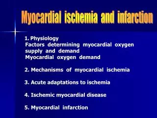
Objectives • To understand the interpretation of 12 lead ECG with regard to • Infarction • Ischemia In the presence of • Right bundle branch block • Left bundle branch block • Ventricular paced rhythms • To understand the utility of decision rules on this topic.
Take-home Messages: • You can make the diagnosis of acute myocardial infarction or ischemia in the face of bundle branch blocks or paced rhythm. • Secondary ST-T wave changes are normal, and go in the opposite direction of the last portion of the QRS complex. • Primary ST-T wave changes mean ischemic or infarction, and go in the same direction as the last portion of the QRS complex.
Take-home Messages: • Left bundle branch block that is new or not known to be old, in the setting of a clinical picture of MI, likely indicates infarction. • Reperfusion therapy is recommended. • Serial ECGs may clarify the situation. • Immediate angiogram is preferred.
Cardiac Conduction System Bachmann’s bundle Sinus node Internodal pathways Left bundle branch AV node Posterior division Bundle of His Anterior division Purkinje fibers Right bundle branch
Truth? • “The diagnosis of myocardial infarction in the presence of left bundle branch block is impossible.”
Partially true… • True: Diagnosis of completed Mi in left bundle branch block is difficult • Q waves may be present with LBBB in the precordial leads without anterior infarction. • ST segment elevation can be hidden in the usual repolarization changes. • But, Q waves in two contiguous lateral leads suggest completed MI • R wave regression from V1-V4 suggests transmural necrosis.
But… • …”ongoing ischemia and injury can be detected in the presence of LBBB, and may be seen as often as they are in the presence of normal cardiac conduction.” • Comparison with old ECGs helpful • Serial ECGs while in the ED also helpful Fesmire, Annals EM, 26:69, 1995
Dr. Braunwald says: • Some findings are highly specific and predictive (90-100%) for MI with left bundle branch block.” • Q waves in at least two contiguous lateral leads (I, aVL, V5 and V6) • R wave regression from V1 to V4 • Primary ST-T wave changes in two or more contiguous leads
ST-T Wave Changes with Bundle Branch Blocks: • Changes are the same with both right and left • Also applies to LVH with “strain” • “Secondary means normal, expected • “Primary” means abnormal: ischemia or infarction
Jpoint ST segment Terminal portion of QRS
Secondary ST-T Wave Changes • These are normal, expected • Terminal portion of the QRS complex is the key • J point displaced away from the terminal portion of the QRS complex • T wave oriented away from the terminal portion of the QRS complex
Primary ST-T Wave Changes • “Primary” = abnormal, not a result of BBB • ST elevation still means injury or infarction • ST depression still means ischemia • Exceptions: • Prinzmetal’s angina: reversible ST elevation • ST depression/T wave inversion can represent infarction: “sub-endocardial” or “non-Q wave.”
Primary ST-T Wave Changes • Must be in two contiguous leads • Inferior: • II and aVF • III and aVF • not II and III • Septal: V1 and V2 • Anterior: V3 and V4 • Lateral: V5, V6, I and aVL (high lateral)
Primary ST-T Wave Changes • One major caveat: • Allowed one lead that has concordant terminal QRS complex and T wave • QRS changes from predominately positive deflection to predominately negative • Don’t infer ischemia/infarction if only one lead
Concept of Dis/Concordance • Refers to whether the last portion of the QRS complex goes in the same or different direction as the T wave • Discordance=good • Concordance=bad
ECG of Evolving MI with Left Bundle Branch Block • Review of 26,003 GUSTO patients (1993) • Derivation set: 131 (0.5%) patients with left bundle branch block • Average time from onset of symptoms to ECG: 120 minutes • Validation set: 45 patients from GUSTO-2A with AMI and LBBB Sgarbossa et al., NEJM, 334:481, 1996
ECG of Evolving MI with Left Bundle Branch Block Identified three predictive criteria: • ST segment elevation > 1 mm concordant with QRS • ST segment depression >1 mm concordant with QRS • ST segment elevation> discordant with QRS • How did these factors perform on the validation set?
ECG of Evolving MI with LBBB • ST elevation > 1 mm concordant with QRS • Sensitivity: 73% • Specificity: 92% • Odds ratio 25.2 (95% CI 11.6-54.7) • ST depression >1 mm concordant with QRS • Sensitivity: 25% • Specificity: 92% • Odds ratio: 6.0 (95% CI 1.9-19.3)
ECG of Evolving MI with Left Bundle Branch Block • ST elevation > 5 mm discordant with QRS • Sensitivity: 26% • Specificity: 92% • Odds ratio: 4.3 (95% CI 1.8-10.6) • Decision tree incorporates all three factors in order of predictive power
ECG of Evolving MI with Left Bundle Branch Block • Does the T wave go the wrong way up? • Does the T wave go the wrong way down? • Does the T wave/ST segment go the right way, but too far? • Three “yes” answers = 100% MI • Three “no” answers = 16% MI
Probability of MI % 100 92 93 88 100 66 50 16
Right Bundle Branch Block in V1 “up down” Secondary normal ST-T Wave changes
Right Bundle Branch Block in V6 “down up” Secondary normal ST-T Wave Changes
Left Bundle Branch Block V1 “down up” Secondary normal ST-T Wave changes
Left Bundle Branch Block V6 “up down” Secondary normal ST-T Wave changes
Right Bundle Branch Block V1 “up up” Primary Infarction ST-T Wave changes
Right Bundle Branch Block V6 “down down” Primary Ischemic ST-T Wave Changes
Left Bundle Branch Block V1 “down down” Primary Ischemic ST-T Wave Changes
Left Bundle Branch Block V6 “up up” Secondary Infarction ST-T Wave Changes
Left Bundle Branch Block V1 > 5 mm “too far up” Primary Infarction ST-T Wave change Exaggerated ST Segment Elevation
Right Ventricular Paced Rhythm with LBBB Pattern: Secondary ST-T Wave Changes
LBBB with Exaggerated ST Elevation: Anteroseptal/lateral Infarction
Left Bundle Branch Block Primary ST T Wave depression Primary STT Wave elevation Exaggerated ST segment elevation
Are These Criteria Valid? • Poor performance • 190 patients/13% with AMI • Sensitivity: 0-16% • Specificity: 93-100% • Treat all LBBB not known to be old as acute MI • Good performance • 224 patients/45% with AMI • Sensitivity: 73% (cardiologist) vs. 67% (EP) • Specificity: 98% (both) Li, et al., Annals EM, 36:561, 2000 Sokolove, et al., Annals EM, 36:566, 2000
ECG of Evolving MI with LBBB • 414 ECGs with AMI and LBBB, 85 with LBBB without AMI • Prevalence of findings: • Concordant ST-segment elevation: 6.3% • Concordant ST-segment depression: 3.1% • Discordant ST-segment elevation: 19.0% • Concordant ST elevation and ST depression in V1-V3 were highly specific for diagnosis of AMI. Gula LJ et al., Coron Artery Dis. 14:387-93, 2003
ECG of LBBB Without MI • 124 patients with LBBB and no MI • Only 1 had primary ST segment depression anteriorly • Only 1 had primary ST segment elevation • 9 had exaggerated ST segment elevation > 5 mm • Sgarbossa criteria are sufficiently specific (few false positives) Madias JE, et al., Clin Cardiol. 24:652-5, 2001
ECG of Evolving MI with LBBB • 182 patients with LBBB and acute MI • New LBBB: Sens = 46%, Spec = 65% • Concordant ST-segment elevation or depression (Sgarbossa criteria) • Specificity = 100% • Positive predictive values = 100% • Sensitivity for ST elevation = 8% • Sensitivity for ST depression = 17% Kontos MC, et al., Ann Emerg Med. 37:431-8, 2001
ECG of Evolving MI with LBBB • Of patients with acute MI and LBBB, the LBBB was NEW (from their MI) in only 46%. • If we only treated NEW LBBB, we’d miss treating 54% of patients who needed it. • Conversely, only 65% of patients with NEW LBBB actually had MI. • So, if we treated everyone with new LBBB and chest pain we’d treat 35% unnecessarily.
Should We Treat All Patients? Yes! • American Heart Association: • Literature: not necessarily • 35% with new LBBB and chest pain did not have an MI (Kontos) • 48% with LBBB not known to be old did not have an MI (Edhouse) • Sgarbossa criteria are helpful • If present, thrombolytics indicated • If absent, serial ECGs or catheterization Edhouse, et al., J Accid Emerg Med. 16:331-5, 1999.
Take-home Messages: • You can make the diagnosis of acute myocardial infarction or ischemia in the face of bundle branch blocks or paced rhythm. • Secondary ST-T wave changes are normal, and go in the opposite direction of the last portion of the QRS complex. • Primary ST-T wave changes mean ischemic or infarction, and go in the same direction as the last portion of the QRS complex.
Take-home Messages: • Left bundle branch block that is new or not known to be old, in the setting of a clinical picture of MI, likely indicates infarction. • Reperfusion therapy is recommended. • Serial ECGs may clarify the situation. • Immediate angiogram is preferred.