Download
1 / 45
460 likes | 779 Vues
Chapter 5: Histology The Study of Tissues. Epithelial Connective Muscle Nerve. Introductory Questions #1. After reading the the introduction on the top of pg. 135, what was the reason for Natalie having a Caesarian section ? What layers of tissue did the physician cut through?

E N D
Chapter 5: HistologyThe Study of Tissues Epithelial Connective Muscle Nerve
Introductory Questions #1 • After reading the the introduction on the top of pg. 135, what was the reason for Natalie having a Caesarian section ? What layers of tissue did the physician cut through? • Name the four major types of tissues that will be examined in this chapter. From these four types of tissues which one would bone and blood be grouped in? • What are some of the distinguishing characteristics of epithelial tissue? • How is epithelial tissue anchored? • What do the following terms mean or indicate: Simple, Stratified, Squamous, Cuboidal, and Columnar
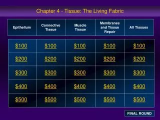
IQ #1 cont’d 6) Matching each type of epithelial lining with its location in the human body. -Intestinal tract A. Simple squamous -Air sacs in the lungs B. Simple cuboidal -Mouth & throat C. Simple columnar -Male urethra D. Pseudostratified columnar -Tubules of the kidney E. Stratified Squamous -Nasal cavities & Resp. tract F. Stratified cuboidal (posseses cilia) G. Stratified columnar -Lines the urinary bladder H. Transitional epithelium http://www.udel.edu/Biology/Wags/histopage/colorpage/colorpage.htm http://www.vetanatomists.org/LIBRARY/histopix.htm http://www.kumc.edu/instruction/medicine/anatomy/histoweb/epithel/epithel.htm http://www.teaching-biomed.man.ac.uk/histology/epith.html
Useful Websites 1.http://www.udel.edu/Biology/Wags/histopage/colorpage/colorpage.htm 2.http://www.vetanatomists.org/LIBRARY/histopix.htm 3.http://www.kumc.edu/instruction/medicine/anatomy/histoweb/epithel/epithel.htm 4. http://www.teaching-biomed.man.ac.uk/histology/epith.html
Epithelial Tissues • Simple squamous • Simple cuboidal • Simple columnar • Pseudostratified columnar (ciliated) • Stratified squamous • Stratified cuboidal • Stratified Columnar • Transitional Epithleium • Glandular Epithelium: (Merocrine, Apocrine, Holocrine)
Functions of Epithelial Tissue • Absorption • Excretion • Protection • Secretion Key terms used to describe and ID epithelial tissues: -Simple: single layer -Stratified: multiple layers -Squamous: thin and flat -Cuboidal: cube-shaped w/large central nucleus -Columnar: elongated cells
Characteristics of Epithelium • Lack blood vessels • Tightly packed • Regenerate frequently • Typical site for developing Cancer Tumors • Forms the lining of internal and external structures and cavities. • Covers most organs • Attached to a nonliving substance called the: Basal lamina (basement membrane) Note**The basal lamina is dissolved away when cancer cells grow.
Simple Squamous Simple Squamous (Pg 136) -alveolar lung sacs -endothelium of heart & blood vessels -cornea of the eye
Simple Cuboidal Simple Cuboidal (See page 137) -forms the lining of the kidney tubules -thyroid gland -lines ducts & glands
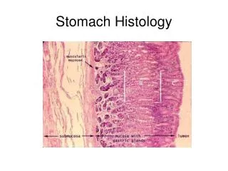
Simple Columnar Simple Columnar (See page 137) Lines the: -Stomach -Intestinal tract (large and small) -Fallopian tubes -Gallbladder Note: notice the nuclei forms a distinct single row
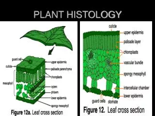
Pseudostratified Columnar Pseudostratified Columnar: (See page 138) -Possesses cilia -Forms a single layer of cells (looks stratified) -Located as the lining for the: *Trachea *Nasal cavities *Bronchi
Stratified Squamous • Lines the: See page 138 • mouth cavity (cheek cells) • throat • vagina • esophagus • skin surface Keratinization occurs on outer layers of the skin -prevents water loss -blocks chemicals from entering -inhibits microorganisms from invading
Stratified Cuboidal • Stratified Cuboidal: See page 139 • Forms the lining of -Ovarian follicles -seminiferous tubules -lumens -larger ducts of glands such as: (mammary, salivary, sweat, and pancreatic)
Stratified Columnar Stratified Columnar: See page 139 • Basal layers consist of cube-shaped cells • Found in the: • Male urethra • Vas deferens • Parts of the pharynx
Transitional Epithelium • See page 140 • Specialized to change in response to increased tension • Creates a barrier to prevent difusion backward • Forms the linings of: • the urinary bladder • passageways of the urinary system
IQ#1 cont’d 6) Matching each type of epithelial lining with its location in the human body. -Intestinal tract A. Simple squamous -Air sacs in the lungs B. Simple cuboidal -Mouth & throat C. Simple columnar -Male urethra D. Pseudostratified columnar -Tubules of the kidney E. Stratified Squamous -Nasal cavities & Resp. tract F. Stratified cuboidal (posseses cilia) G. Stratified columnar -Lines the urinary bladder H. Transitional epithelium
Introductory Questions #2 • Exocrine glands have six different structural types. Name them. • How do Merocrine glands, Apocrine glands, and Holocrine glands differ? Where is each gland located? • How is serous fluid different from mucous? • A unicellular gland is also called a(n) gland. • How does connective tissue differ from epithelial tissue? • What are the major types of cells that comprise connective tissue?
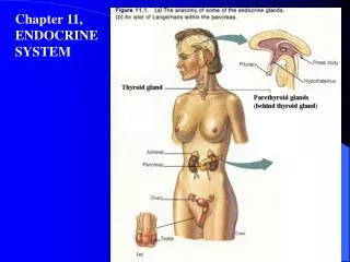
Glandular Epitheliumhttp://www.med.uiuc.edu/histo/large/atlas/search.htm • Cells specialized to produce and secrete substances into ducts or into body fluids. • 2 types: Exocrine and Endocrine • Characterized by what they release and what they release it into. • Endocrine Glands: secretes into blood or tissue • Ex. Hormones (Ch. 13) • Six structural types of exocrine glands (pg 141) • Simple exocrine glands: four types • Compound exocrine glands: two types
Introductory Questions #3 Matching: • Mammary A. Apocrine • Sebaceous gland B. Merocrine • Ceruminous C. Holocrine • Pancreatic glands • Salivary glands • Composed of Serous or mucous cells
Connective Tissue • Characteristics: -Good blood supply -Cells spaced farther apart -Occurs throughout the body (most mass) • Functions: -Bind -Fill spaces -Support -Store fat -Protect -Prod. Blood cells
Cell types of Connective Tissue • Resident Cells: present in stable numbers Ex. Fibroblasts & Mast cells • Wandering Cells: appears temporarily Ex. Macrophage & Leukocytes (WBC’s)
List of Cell Types • Fibroblasts Produces 3 types of Fibers • Mast Cells Inflammation & Prev. clotting • Macrophages Phagocytic Cells • Adipocytes Fat • Chondrocytes Cartilage • Osteocytes Bone • Erythrocytes & Leukocytes: Blood
Fibroblasts • Star-shaped cell • Secretes protein to produce fibers • 3 Types of fibers: • Collagenous: thick, slightly elastic • Elastic: Microfibrils w/ elastin protein • Reticular: very thin fibers, highly branched *Strength: collagenouselasticreticular
Collagenous Fibers • Collagenous: -Very strong, great tensile strength -long, parallel bundles -holds bones together Ex. ligaments & tendons Many = Dense Connective Tissue (White) Sparse = Loose Connective Tissue
Elastic Fibers • Composed of Microfibrils • Protein is called elastin • Stretches and resumes back to original length • Called “Yellow fibers” • Common in areas of frequent stretching Ex. Vocal cords, walls of hollow organs, airways vertebrae, walls of heart and larger Arteries
Muscle Tissue • Skeletal Muscle • Cardiac Muscle • Smooth Muscle (nonstriated)