Download
1 / 77
780 likes | 896 Vues
Citric acid cycle supplies NADH and FADH 2 to the electron transport chain. Fatty Acids. Acetyl Co A. Amino Acids. Pyruvate. Glucose. Reduced coenzymes NADH and FADH 2 are formed in matrix from: (1) Oxidative decarboxilation of pyruvate to acetyl CoA

E N D
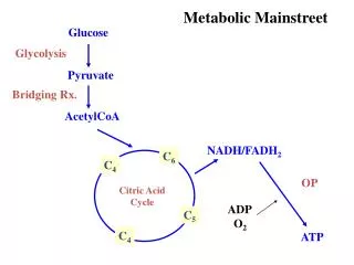
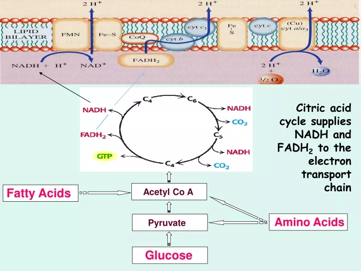
Citric acid cycle supplies NADH and FADH2 to the electron transport chain Fatty Acids Acetyl Co A Amino Acids Pyruvate Glucose
Reduced coenzymes NADH and FADH2 are formed in matrix from: (1) Oxidative decarboxilation of pyruvate to acetyl CoA (2) Aerobic oxidation of acetyl CoA by the citric acid cycle (3) Oxidation of fatty acids and amino acids The NADH and FADH2 are energy-rich molecules because each contains a pair of electrons having a high transfer potential.
Electrons of NADH or FADH2 are used to reduce molecular oxygen to water. A large amount of free energy is liberated. The electrons from NADH and FADH2are not transported directly to O2 but are transferred through series of electron carriers that undergo reversible reduction and oxidation.
The flow of electrons through carriers leads to the pumping of protons out of the mitochondrial matrix. The resulting distribution of protons generates a pH gradient and a transmembrane electrical potential that creates a protonmotive force.
ATP is synthesized when protons flow back to the mitochondrial matrix through an enzyme complex ATP synthase. The oxidation of fuels and the phosphorylation of ADP are coupled by a proton gradient across the inner mitochondrial membrane. Oxidative phosphorylation is the process in which ATP is formed as a result of the transfer of electrons from NADH or FADH2 to O2 by a series of electron carriers.
OXIDATIVE PHOSPHORYLATION IN EUKARYOTES TAKES PLACE IN MITOCHONDRIA Two membranes: outer membrane inner membrane (folded into cristae) Two compartments: (1) the intermembrane space (2) the matrix Location of mitochondrial complexes • Inner mitochondrial membrane: Electron transport chainATP synthase • Mitochondrial matrix:Pyruvate dehydrogenase complexCitric acid cycleFatty acid oxidation The outer membrane is permeable to small molecules and ions because it contains pore-forming protein (porin). The inner membrane is impermeable to ions and polar molecules. Contains transporters (translocases).
NADH FMN Fe-S Co-Q Fe-S cyt c1 cyt c cyt a cyt a3 O2 cyt b succinate FAD Fe-S THE ELECTRON TRANSPORT CHAIN Series of enzyme complexes (electron carriers) embedded in the inner mitochondrial membrane, which oxidize NADH2 and FADH2 and transport electrons to oxygen is calledrespiratory electron-transport chain (ETC). The sequence of electron carriers in ETC
High-Energy Electrons: Redox Potentials and Free-Energy Changes In oxidative phosphorylation, the electron transfer potentialof NADH or FADH2 is converted into the phosphoryl transfer potentialof ATP. Phosphoryl transfer potential is G°' (energy released during the hydrolysis of activated phos-phate compound). G°' for ATP = -7.3 kcal mol-1 Electron transfer potential is expressed as E'o, the (also called redox potential, reduction potential, or oxidation-reduction potential).
NADH FMN Fe-S Co-Q Fe-S cyt c1 cyt c cyt a cyt a3 O2 cyt b succinate FAD Fe-S THE RESPIRATORY CHAIN CONSISTS OF FOUR COMPLEXES I II Components of electron-transport chain are arranged in the inner membrane of mitochondria in packages called respiratory assemblies (complexes). III IV III IV I II
Complex I (NADH-ubiquinone oxidoreductase) Transfers electrons from NADH to Co Q (ubiquinone)Consist of: - enzyme NADH dehydrogenase(FMN - prosthetic group) - iron-sulfur clusters. NADH reduces FMN to FMNH2. Electrons from FMNH2 pass to a Fe-S clusters. Fe-S proteins convey electrons to ubiquinone. QH2 is formed. The flow of two electrons from NADH to coenzym Q leads to the pumping of four hydrogen ions out of the matrix.
Complex II (succinate-ubiquinon oxidoreductase) Transfers electrons from succinate to Co Q.Form 1 consist of: - enzyme succinate dehydrogenase (FAD – prosthetic group) - iron-sulfur clusters. Succinate reduces FAD to FADH2. Then electrons pass to Fe-S proteins which reduce Q to QH2 Form 2 and 3 contains enzymes acyl-CoA dehydrogenase (oxidation of fatty acids) and glycerol phosphate dehydrogenase (oxidation of glycerol) which direct the transfer of electrons from acyl CoA to Fe-S proteins. Complex II does not contribute to proton gradient.
All electrons must pass through the ubiquinone (Q)-ubiquinole (QH2) pair. Ubiquinone Q:- lipid soluble molecule, - smallest and most hydrophobic of all the carriers - diffuses within the lipid bilayer - accepts electrons from I and II complexes and passes them to complex III.
Complex III (ubiquinol-cytochrome c oxidoreductase) Transfers electrons from ubiquinol to cytochrome c.Consist of:cytochrome b, Fe-S clusters and cytochrome c1.Cytochromes – electron transferring proteins containing a heme prosthetic group (Fe2+ Fe3+). Oxidation of one QH2 is accompanied by the translocation of 4 H+ across the inner mitochondrial membrane. Two H+ are from the matrix, two from QH2
Complex IV (cytochrome c oxidase) Transfers electrons from cytochrome c to O2.Composed of:cytochromes a and a3. Catalyzes a four-electron reduction of molecular oxygen (O2) to water (H2O): O2 + 4e- + 4H+ 2H2O Translocates 2H+ into the intermembrane space
A PROTON GRADIENT POWERS THE SYNTHESIS OF ATP The transport of electrons from NADH or FADH2 to O2 via the electron-transport chain is exergonic process: NADH + ½O2 + H+ H2O + NAD+ FADH2+½O2 H2O + FAD+ Go’ = -52.6 kcal/mol for NADH -36.3 kcal/mol for FADH2 How this process is coupled to the synthesis of ATP (endergonic process)?ADP + Pi ATP + H2OGo’=+7.3 kcal/mol
The Chemiosmotic Theory • Proposed by Peter Mitchell in the 1960’s (Nobel Prize, 1978) • Chemiosmotic theory: electron transport and ATP synthesis are coupled by a proton gradient across the inner mitochondrial membrane Mitchell’s postulates for chemiosmotic theory • Intact inner mitochondrial membrane is required • Electron transport through the ETC generates a proton gradient • 3. ATP synthase catalyzes the phosphorylation of ADP in a reaction driven by movement of H+ across the inner membrane into the matrix
REGULATION OF OXIDATIVE PHOSPHORYLATION Coupling of Electron Transport with ATP Synthesis Electron transport is tightly coupled to phosphorylation. ATP can not be synthesized by oxidative phosphorylation unless there is energy from electron transport. Electrons do not flow through the electron-transport chain to O2 unless ADP is phosphorylated to ATP. Important substrates: NADH, O2, ADP Intramitochondrial ratio ATP/ADPis a control mechanism High ratio inhibits oxidative phosphorylation as ATP allosterically binds to a subunit of Complex IV
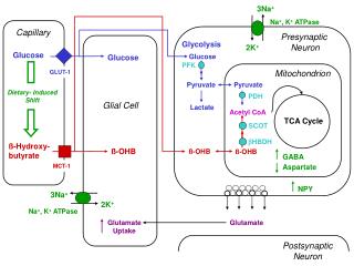
Uncoupling of Electron Transport with ATP Synthesis Uncoupling of oxidative phosphorylation generates heat to maintain body temperature in hibernating animals, in newborns, and in mammals adapted to cold. Brown adipose tissues is specialized for thermogenesis. Inner mitochondrial membrane contains uncoupling protein (UCP), or thermogenin. UCP forms a pathway for the flow of protons from the cytosol to the matrix.
Uncouplers • Uncouplers are lipid-soluble aromatic weak acids • Uncouplers deplete proton gradient by transporting protons across the membrane 2,4-Dinitrophenol: an uncoupler • Because the negative charge is delocalized over the ring, both the acid and base forms of DNP are hydrophobic enough to dissolve in the membrane.
Specific inhibitors of electron transport chain and ATP-synthase Specific inhibitors of electron transport are invaluable in revealing the sequence of electron carriers. Rotenoneand amytalblock electron transfer in Complex I. Antimycin Ainterferes with electron flow thhrough Complex III. Cyanide, azide,and carbon monoxideblock electron flow in Complex IV. ATP synthaseis inhibitedbyoligomycin which prevent the influx of protons through ATP synthase.
3 4 4 2 ATP Yield Ten protons are pumped out of the matrix during the two electrons flowing from NADH to O2 (Complex I, III and IV). Six protons are pumped out of the matrix during the two electrons flowing from FADH2 to O2 (Complex III and IV). Translocation of 3H+ required by ATP synthase for eachATP produced 1 H+ needed for transport of Pi. Net:4 H+ transported for each ATP synthesized For NADH: 10 H+/ 4H+) = 2.5 ATP For FADH2: 6 H+/ 4 H+ = 1.5 ATP
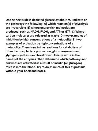
DIGESTION OF CARBOHYDRATES Glycogen, starch and disaccharides(sucrose, lactose and maltose) are hydrolyzed to monosaccharide units in the gastrointestinal tract. The process of digestion starts in the mouth by the salivary enzyme –amilase. The time for digestion in mouth is limited. Salivary -amilaseis inhibited in stomach due to the action of hydrochloric acid. Another -amilaseis produced in pancreas and is available in the intestine.
-amilase -amilase hydrolyzes the -1-4-glycosidic bonds randomlyto produce smaller subunits like maltose, dextrines and unbranched oligosaccharides.
sucrase The intestinal juice contains enzymes hydrolyzing disaccharides into monosaccharides (they are produced in the intestinal wall) Sucrase hydrolyses sucrose into glucose and fructose Glucose Fructose Sucrose
lactase maltase Maltose Glucose Galactose Lactase hydrolyses lactose into glucose and galactose Lactose Glucose Glucose Maltase hydrolyses maltose into two glucose molecules
Protein Na+ Glucose ABSORPTION OF CARBOHYDRATES Only monosaccharides are absorbed The rate of absorption: galactose > glucose > fructose Glucose and galactose from the intestine into endothelial cells are absorbed by secondary active transport Protein
Transport of glucose from blood into cells of different organs is mainly by facilitated diffusion. The protein facilitating the glucose transport is called glucose transporter (GluT). GluT are of 5 types. GluT2 is located mainly in hepatocytes membranes (it transport glucose into cells when blood sugar is high); GluT1 is seen in erythrocytes and endothelial cells; GluT3 is located in neuronal cells (has higher affinity to glucose); GluT5 – in intestine and kidneys; GluT4 - in muscles and fat cells.
The fate of glucose molecule in the cell Glucose Pentose phosphate pathway supplies the NADPH for lipid synthesis and pentoses for nucleic acid synthesis Glycogenogenesis (synthesis of glycogen) is activated in well fed, resting state Glucose-6-phosphate Ribose, NADPH Glycogen Pyruvate Glycolysis is activated if energy is required
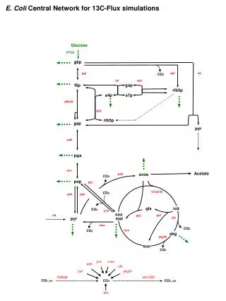
Catabolism of glucose in aerobic conditions via glycolysis and the citric acid cycle
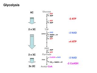
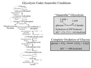
Glycolysis (10 reactions) can be divided into three stages • In the 1st stage (hexosestage) 2 ATP are consumed per glucose • In the 3rd stage (triosestage)4 ATP are produced per glucose • Net: 2 ATP produced per glucose
Stage 1, which is the conversion of glucose into fructose 1,6-bisphosphate, consists of three steps: a phosphorylation, an isomerization, and a second phosphorylation reaction. The strategy of these initial steps in glycolysis is to trap the glucose in the cell and form a compound that can be readily cleaved into phospho-rylated three-carbon units.
Stage 2 is the cleavage of the fructose 1,6-bisphosphate into two three-carbon fragments dihydroxyacetone phosphate and glyceraldehyde 3-phosphate. Dihydroxyacetone phosphate and glyceraldehyde 3-phosphate are readily interconvertible.
In stage 3, ATP is harvested when the three-carbon fragments are oxidized to pyruvate.
1. Hexokinase Glycolysis Has 10 Enzyme-Catalyzed Steps • Each chemical reaction prepares a substrate for the next step in the process • Transfers the g-phosphoryl of ATP to glucose C-6 oxygen to generate glucose 6-phosphate (G6P) • Four kinases in glycolysis: steps 1,3,7, and 10 • All four kinases require Mg2+ and have a similar mechanism
2. Glucose 6-Phosphate Isomerase • Converts glucose 6-phosphate (G6P) (an aldose) to fructose 6-phosphate (F6P) (a ketose) • Enzyme preferentially binds the a-anomer of G6P (converts to open chain form in the active site) • Enzyme is highly stereospecific for G6P and F6P • Isomerase reaction is near-equilibrium in cells
3. Phosphofructokinase-1 (PFK-1) • Catalyzes transfer of a phosphoryl group from ATP to the C-1 hydroxyl group of F6P to form fructose 1,6-bisphosphate (F1,6BP) • PFK-1 is metabolically irreversible and a critical regulatory point for glycolysis in most cells • A second phosphofructokinase (PFK-2) synthesizes fructose 2,6-bisphosphate (F2,6BP)
4. Aldolase • Aldolase cleaves the hexose F1,6BP into two triose phosphates: glyceraldehyde 3-phosphate (GAP) and dihydroxyacetone phosphate (DHAP) • Reaction is near-equilibrium, not a control point
5. Triose Phosphate Isomerase (TPI) • Conversion of DHAP into GAP • Reaction is very fast, only the D-isomer of GAP is formed • Reaction is reversible. At equilibrium, 96% of the triose phosphate is DHAP. However, the reaction proceeds readily from DHAP to GAP because the subsequent reactions of glycolysis remove this product.
6. Glyceraldehyde 3-Phosphate Dehydrogenase (GAPDH) • Conversion of GAP to 1,3-bisphosphoglycerate (1,3BPG) • Molecule of NAD+ is reduced to NADH • Energy from oxidation of GAP is conserved in acid-anhydride linkage of 1,3BPG • Next step of glycolysis uses the high-energy phosphate of 1,3BPG to form ATP from ADP
7. Phosphoglycerate Kinase (PGK) • Transfer of phosphoryl group from the energy-rich mixed anhydride 1,3BPG to ADP yields ATP and 3-phosphoglycerate (3PG) • Substrate-level phosphorylation - Steps 6 and 7 couple oxidation of an aldehyde to a carboxylic acid with the phosphorylation of ADP to ATP
8. Phosphoglycerate Mutase • Catalyzes transfer of a phosphoryl group from one part of a substrate molecule to another • Reaction occurs without input of ATP energy
9. Enolase: 2PG to PEP • 2-Phosphoglycerate (2PG) is dehydrated to phosphoenolpyruvate (PEP) • Elimination of water from C-2 and C-3 yields the enol-phosphate PEP • PEP has a veryhigh phosphoryl group transfer potential because it exists in its unstable enol form
10. Pyruvate Kinase (PK) PEP + ADP Pyruvate + ATP • Catalyzes a substrate-level phosphorylation • Metabolically irreversible reaction • Regulation both by allostericmodulators and by covalentmodification • Pyruvate kinase gene can be regulated by various hormones and nutrients
Net reaction of glycolysis During the convertion of glucose to pyruvate: • Two molecules of ATP are produced • Two molecules of NAD+ are reduced to NADH Glucose + 2 ADP + 2 NAD+ + 2 Pi 2 Pyruvate + 2 ATP + 2 NADH + 2 H+ + 2 H2O
The Fate of Pyruvate The sequence of reactions from glucose to pyruvate is similar in most organisms and most types of cells. The fate of pyruvate is variable. Three reactions of pyruvate are of prime importance: 1. Aerobic conditions: oxidation to acetyl CoA which enters the citric acid cycle for further oxidation 2. Anaerobic conditions(muscles, red blood cells): conversion tolactate 3. Anaerobic conditions (microorganisms, yeast):conversion to ethanol
Metabolism of Pyruvate to Ethanol Ethanolis formed from pyruvate in yeast andseveral other microorganisms in anaerobic conditions. Two reactions required: The first step is the decarboxylation of pyruvate to acetaldehyde. Enzyme - pyruvate decarboxylase. Coenzyme - thiamine pyrophosphate (derivative of the vitamin thiamine B1) The second step is the reduction of acetaldehyde to ethanol. Enzyme - alcohol dehydrogenase(active site contains a zinc). Coenzyme – NADH.
The conversion of glucose into ethanol is an example of alcoholic fermentation. The net result of alcoholic fermentation is: Glucose+2Pi + 2ADP + 2H+ 2ethanol + 2CO2 + 2ATP + 2H2O The ethanol formed in alcoholic fermentation provides a key ingredient for brewing and winemaking. There is no net NADH formation in the conversion of glucose into ethanol. NADH generated by the oxidation of glyceraldehyde 3-phosphate is consumed in the reduction of acetaldehyde to ethanol.
Metabolism of Pyruvate to Lactate Lactateis formed from pyruvate in an animal organism and in a variety of microorganisms in anaerobic conditions. The conversion of glucose into lactate is called lactic acid fermentation. Enzyme - lactate dehydrogenase. Coenzyme – NADH.
Muscles of higher organisms and humans lack pyruvate decarboxylase and cannot produce ethanol from pyruvate • Muscle contain lactate dehydrogenase. During intense activity when the amount of oxygen is limiting the lactic acid can be accumulated in muscles (lactic acidosis). • Lactate formed in skeletal muscles during exercise is transported to the liver. • Liver lactate dehydrogenase can reconvert lactate to pyruvate. • Overall reaction in the conversion of glucose into lactate: • Glucose + 2 Pi + 2 ADP 2lactate + 2 ATP + 2 H2O • As in alcoholic fermentation, there is no net NADH formation. • NADH formed in the oxidation of glyceraldehyde 3-phosphate is consumed in the reduction of pyruvate.