Download
1 / 54
540 likes | 601 Vues
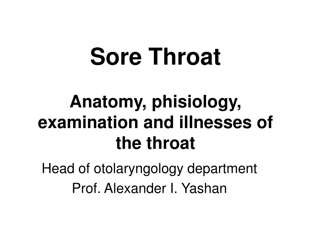
Sore Throat Anatomy, phisiology, examination and illnesses of the throat. Head of otolaryngology department Prof. Alexander I. Yashan. Anatomy of the nose. PHISIOLOGY of the NOSE. Breathing Smelling Protection Cosmetics Speech (articulation). Protection. Warming Cleaning Moisten

E N D
Sore ThroatAnatomy, phisiology, examination and illnesses of the throat Head of otolaryngology department Prof. Alexander I. Yashan
PHISIOLOGY of the NOSE • Breathing • Smelling • Protection • Cosmetics • Speech (articulation)
Protection • Warming • Cleaning • Moisten • Buffering
Bony shelves in the nasal cavity Inferior, middle and superior (at times, supreme) Airflow and defense mechanism Only inferior and middle turbinates are seen with speculum Examined for colour, masses, discharge, swelling and lesions Turbinates
Part of middle meatues where maxillary, frontal and anterior ethmoid sinuses drain Any deviation and disease can obstruct natural drainange pathways Swollen turbinates with inflammed mucosa often indicate unlerlying diseases Middle Turbinate
Easily visible via speculum view Examination is often under headlight Enlargement of turbinate can cause chrosnic nasal obstruction Bone can thickens and mucosa can swell to narrow nasal airspace Inferior Turbinate
Sinuses are Lined with Cilia Which Beat to Transport Mucous Through and Out of the Sinuses Bacterial Infection Can Cause Changes to Occur Including Swelling of the Mucousal Lining Cilia Cease to Function Properly Ostium Closes trapping Mucous Inside Sinus Cavity Sinus Mucosa
Drainage of the maxillary,ethmoid and frontal sinuses via their ostia and very narrow chambers before entering the middle meatus - ethmoidal infindibulum and the frontal recess Bacterial infection, allergies, polyps etc. can cause swelling and stenosis of these very narrow channels Ethmoidal Prechambers
PHISIOLOGY of the THROAT • Breathing • Swallowing • Separating (channelization) • Speech (articulation)
SWALLOWING • Normal mechanism - 3 stages • 1st Stage - Oral (Voluntary) - tongue pushed against palate, forcing food into pharynx, triggering reflex stages • 2nd Stage - Pharyngeal involuntary lasts 1-2 seconds Food in pharynx stimulates receptors with afferents in V and IX leading to the medulla. Reflex efferent signals travel via V, IX, X, and XII to: • Elevate soft palate to seal off nasopharynx • Move palatopharyngeal walls medially • Close glottis and depress epiglottis • Larynx moves superiorly, and anteriorly under base of tongue to shield larynx and widen hypopharynx • Relax cricopharyngeus • Close superior constrictor as bolus passes into esophagus • 3rd Stage - Esophageal (Involuntary) • Liquids usually fall by gravity • Peristaltic waves push solids. Innervated by vagi and myenteric plexus.
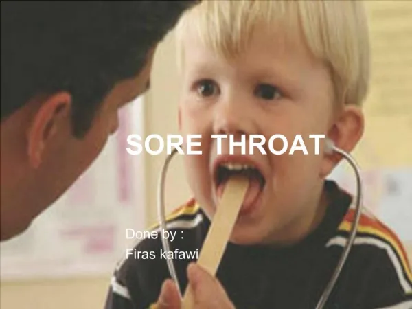
Examination Scheme External: Lips Oral vestibule Teeth and gums Hard & soft palate Palatal mobility Tongue dorsal, ventral surfaces, Floor of mouth Tongue mobility Put Tongue Depressor & examine: Tonsils Ant. & post. Pillars Tongue Posterior 1/3 Post. Pharyngeal wall & its mobility
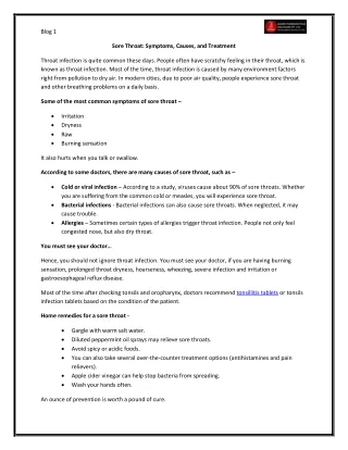
INFLAMMATORY DISORDERS OF THE PHARYNX • Inflammatory disorders of the pharynx most commonly present as throat or neck pain. Disphagia, odynophagia, and airway obstruction are other frequent complaints. The pharynx is a dynamic conduit for inspired air and ingested matter, responsible for diverting each into the trachea or esophagus, respectively. This process may be impaired by anything which obstructs or restricts the mobility of the pharynx. The following outline is directed toward a systematic approach to the evaluation of the patient with sore throat, odynophagia or disphagia.
EVALUATION • Key historical considerations • Age of patient • Onset and duration • History of recent trauma (including possible foreign body) • Inflammatory symptoms - fever, pain, malaise, malodorous breath • Status of nasal airway: congestion, obstruction, rhinorrhea, purulent discharge, allergic history, snoring • Reflux symptoms such as heartburn or water brash • Associated ear pain • Disphagia or odynophagia • Dyspnea or stridor • Other associated symptoms • Recent exposure to infectious discharge • Cancer risk factors: smoking history, ETOH abuse
Key considerations of physical examination for patients with throat pain: • Ears - The patient's ears need to be examined for primary ear pathology, as acute otitis media and serous otitis media are often preceded by pharyngitis and nasal congestion. Conversely many patients with pharyngeal inflammation or tumor will have referred ear pain in which case otoscopy will be normal. • Nose - The nose should be examined for any evidence of obstruction, purulence, or excessive secretions. Mouth breathing leads to drying of pharyngeal mucosa; this is a very common cause of chronic sore throat. Excessive secretion may cause the patient to clear his throat frequently, which traumatizes the larynx; and infected drainage from sinusitis may cause irritation in the pharynx. • Pharynx - Examination of the throat for asymmetry, injection, erythema, exudate, swelling, or pooling of secretions. Also, careful inspection and palpation of any ulcerations, lesions, mucosal or submucosal masses. • Neck - Careful palpation and inspection of the neck for lymphadenopathy, swelling, tenderness, induration or fluctuance. Large, firm, non-tender masses suggest neoplasia, while multiple small nodes are often seen in chronic recurrent infections.
Acute Viral or Bacterial Pharyngitis • Pharyngitis is caused by a variety of microorganisms. Most cases are viral and include the virus causing the common cold, flu (influenza virus), adenovirus, mononucleosis, HIV among various others. Bacterial causes include Group A streptococcus which causes strep throat (15% of cases), in addition to Corynebacterium, Arcanobacterium, Neisseria gonorrhoeae, Chlamydia pneumoniae and others. In up to 30% of cases, no organism is identified. • Most cases of pharyngitis occur during the colder months -- during respiratory disease season. Spread among household members is common. The medical importance of recognizing strep throat as a cause of pharyngitis stems from the need to prevent its complications which can include acute rheumatic fever, kidney dysfunction and severe disease such as bacteremia and rarely streptococcal toxic shock syndrome. http://weed.ru/d/
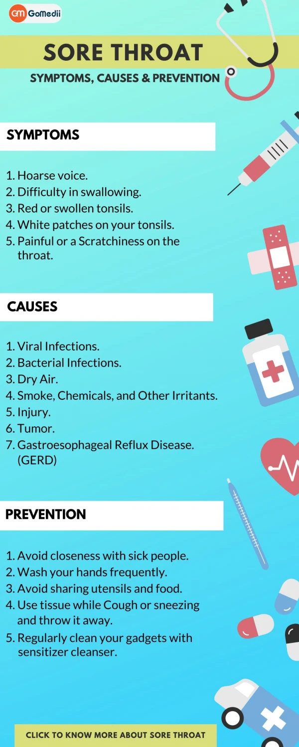
Symptoms • sore throat • additional symptoms are dependent on the underlying microorganisms • step throat may be accompanied by fever, headache, swollen lymph nodes in the neck • viral pharyngitis may be associated with runny nose (rhinorrhea) and postnasal discharge • severe cases of pharyngitis may be accompanied by difficulty swallowing and rarely difficulty breathing • Signs and tests A physical exam with attention to the pharynx to assess whether drainage/coating (exudates) are present, as well as skin, eyes, neck lymph nodes is frequently done.
Oropharyngoscopy • Swollen, erythematous mucosa of the oropharynx and hypopharynx, often with edema of the uvula and soft palate. • Swollen cyanotic lymphatic follicles on the posterior wall • Mucous or purulent discharges on the posterior wall
Complications • complications of strep throat: • rheumatic fever, • glomerulonephritis (kidney inflammation), • chorea, • bacteremia (bloodstream infection) and rarely streptococcal shock syndrome • in some severe forms of pharyngitis (e.g., severe mononucleosis-pharyngitis) • airway obstruction may occur • peritonsillar abscess, retropharyngeal abscess
Acute Tonsillitis • The most common organism is beta hemolytic streptococcus, but viral organisms can also cause exudative tonsillitis. Other causative organisms include staphylococcus aureus, streptococcus viridans, and various hemophilus species.
General Symptoms • Rapid onset of throat pain with pain on swallowing associated with • Fever, often 38°-39° C • Malaise • fatigue • Chill • Pain in extremities, muscles and joints
Catharal and Follicular Tonsilitis • The tonsils are red, enlarged and painfulness • with an exudate or studded with white follicles. • Tender, firm cervical adenopathy is often present.
Adenotomy • Adenoid grades
Asymmetrical tonsils Large kissing tonsils
In mononucleosis the tonsils are hyperaemic and pusaccumulates in the tonsillar crypts. The debris in the crypts coalescesto form a purulent membrane. The clinical picture resemblesof that in streptococcal tonsillitis
Right peritonsillar abscess; the peritonsillar space, thesoft palate and the uvula are swollen. The uvula is displaced tothe contralateral side
Keratosis Concretions, exudate
Tonsil Tumours Carcinoma Papilloma