Download
1 / 66
660 likes | 667 Vues
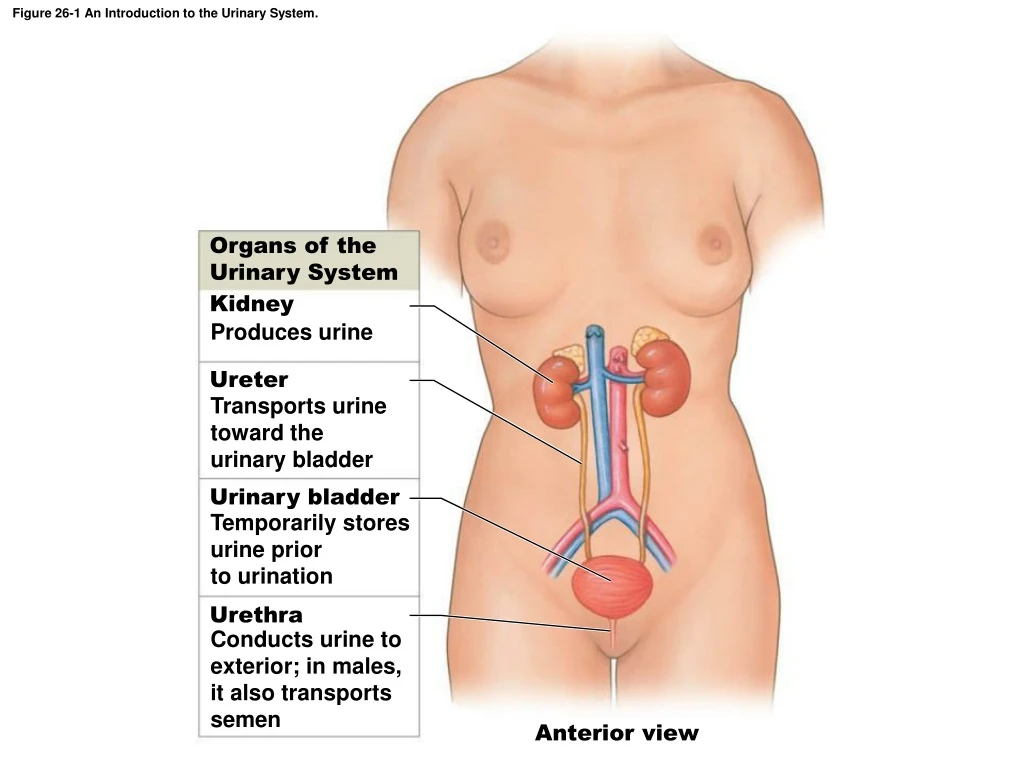
Organs of the Urinary System. Figure 26-1 An Introduction to the Urinary System. Kidney. Produces urine. Ureter. Transports urine toward the urinary bladder. Urinary bladder. Temporarily stores urine prior to urination. Urethra. Conducts urine to exterior; in males,

E N D
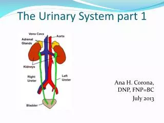
Organs of the Urinary System Figure 26-1 An Introduction to the Urinary System. Kidney Produces urine Ureter Transports urine toward the urinary bladder Urinary bladder Temporarily stores urine prior to urination Urethra Conducts urine to exterior; in males, it also transports semen Anterior view
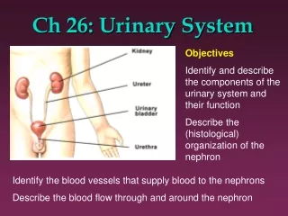
26-1 Urinary System Functions • Three Functions of the Urinary System • Excretion • Removal of organic wastes from body fluids • Elimination • Discharge of waste products • Homeostatic regulation • Of blood plasma volume and solute concentration
Cortical radiate veins Cortical radiate arteries Interlobar arteries Cortex Segmental artery Figure 26-5a The Blood Supply to the Kidneys. Adrenal artery Renal artery Renal vein Arcuate veins Interlobar veins Medulla Arcuate arteries A sectional view, showing major arteries and veins a
Glomerulus Cortical radiate vein Afferent arterioles Cortical radiate artery Arcuate artery Figure 26-5b The Blood Supply to the Kidneys. Cortical nephron Arcuate vein Juxtamedullary nephron Renal pyramid Interlobar vein Interlobar artery Minor calyx Circulation in a single kidney lobe b
NEPHRON Proximal convoluted tubule Distal convoluted tubule • Secretion of ions, acids, drugs, toxins • Variable reabsorption of water, sodium ions, and calcium ions (under hormonal control) • Reabsorption of water, ions, and all organic nutrients Cuboidal cells with abundant microvilli Cuboidal cells with few microvilli Mitochondria Renal tubule Renal corpuscle Figure 26-6 The Functional Anatomy of a Representative Nephron and the Collecting System (Part 1 of 2). • Production of filtrate Squamous cells Efferent arteriole Afferent arteriole Glomerulus Glomerular capsule Ascending limb of loop ends Descending limb of loop begins Capsular space Nephron loop Descending limb Further reabsorption of water Thick ascending limb Squamous cells Ascending limb Reabsorption of sodium and chloride ions Thin descending limb Low cuboidal cells KEY Solute reabsorption or secretion Filtrate Water reabsorption Variable solute reabsorption or secretion Variable water reabsorption
COLLECTING SYSTEM Collecting duct • Variable reabsorption of water and reabsorption or secretion of sodium, potassium, hydrogen, and bicarbonate ions Figure 26-6 The Functional Anatomy of a Representative Nephron and the Collecting System (Part 2 of 2). Intercalated cell Principal cell KEY Papillary duct Filtrate Water reabsorption • Delivery of urine to minor calyx Variable water reabsorption Solute reabsorption or secretion Columnar cells Minor calyx Variable solute reabsorption or secretion
26-2 The Kidneys • Cortical Nephrons • 85 percent of all nephrons • Located mostly within superficial cortex of kidney • Nephron loop (Loop of Henle) is relatively short • Efferent arteriole delivers blood to a network of peritubular capillaries • Juxtamedullary Nephrons • 15 percent of nephrons • Nephron loops extend deep into medulla • Peritubular capillaries connect to vasa recta
Cortical nephron Juxtamedullary nephron Cortex Figure 26-7a The Locations and Structures of Cortical and Juxtamedullary Nephrons. Medulla Collecting duct Papillary duct Renal papilla Minor calyx The general appearance and location of nephrons in the kidneys a
Distal convoluted tubule Peritubular capillaries Efferent arteriole Afferent arteriole Figure 26-7b The Locations and Structures of Cortical and Juxtamedullary Nephrons. Renal corpuscle Collecting duct Peritubular capillaries Nephron loop The circulation to a cortical nephron b
Peritubular capillaries Distal convoluted tubule (DCT) Proximal convoluted tubule (PCT) Figure 26-7c The Locations and Structures of Cortical and Juxtamedullary Nephrons. Renal corpuscle Collecting duct Vasa recta Nephron loop The circulation to a juxtamedullary nephron c
NEPHRON Proximal convoluted tubule Distal convoluted tubule • Secretion of ions, acids, drugs, toxins • Variable reabsorption of water, sodium ions, and calcium ions (under hormonal control) • Reabsorption of water, ions, and all organic nutrients Cuboidal cells with abundant microvilli Cuboidal cells with few microvilli Mitochondria Renal tubule Renal corpuscle Figure 26-6 The Functional Anatomy of a Representative Nephron and the Collecting System (Part 1 of 2). • Production of filtrate Squamous cells Efferent arteriole Afferent arteriole Glomerulus Glomerular capsule Ascending limb of loop ends Descending limb of loop begins Capsular space Nephron loop Descending limb Further reabsorption of water Thick ascending limb Squamous cells Ascending limb Reabsorption of sodium and chloride ions Thin descending limb Low cuboidal cells KEY Solute reabsorption or secretion Filtrate Water reabsorption Variable solute reabsorption or secretion Variable water reabsorption
Glomerular capsule Capsular epithelium Visceral epithelium (podocyte) Glomerular capillary Capsular space Figure 26-8a The Renal Corpuscle. Efferent arteriole Proximal convoluted tubule Distal convoluted tubule Juxtaglomerular complex Macula densa Juxtaglomerular cells Afferent arteriole Important structural features of a renal corpuscle. a
Filtration membrane Podocyte nucleus Fenestrated endothelium Dense layer Filtration slits Figure 26-8b The Renal Corpuscle. Capillary endothelial cell Mesangial cell Pores RBC Pedicels Podocyte Capsular space Capsular epithelium This cross section through a portion of the glomerulus shows the components of the filtration membrane of the nephron. b
26-2 The Kidneys • The Glomerular Capillaries • Are fenestrated capillaries • Endothelium contains large-diameter pores • Blood Flow Control • Special supporting cells (mesangial cells) • Between adjacent capillaries • Control diameter and rate of capillary blood flow
26-2 The Kidneys • The Filtration Membrane • Consists of: • Fenestrated endothelium • Dense layer • Filtration slits
26-2 The Kidneys • The Juxtaglomerular Complex (JGC) • An endocrine structure that secretes: • Hormone erythropoietin • Enzyme renin • Formed by: • Macula densa • Juxtaglomerular cells
26-2 The Kidneys • Macula Densa • Epithelial cells of DCT, near renal corpuscle • Tall cells with densely clustered nuclei • Juxtaglomerular Cells • Smooth muscle fibers in wall of afferent arteriole • Associated with cells of macula densa • Together with macula densa forms juxtaglomerular complex (JGC)
26-2 The Kidneys • Filtration • Blood pressure • Forces water and small solutes across membrane into capsular space • Larger solutes, such as plasma proteins, are excluded
26-2 The Kidneys • Filtration at Renal Corpuscle • Is passive • Solutes enter capsular space • Metabolic wastes and excess ions • Glucose, free fatty acids, amino acids, and vitamins • Reabsorption • Useful materials are recaptured before filtrate leaves kidneys • Reabsorption occurs in proximal convoluted tubule
26-2 The Kidneys • The Proximal Convoluted Tubule (PCT) • Is the first segment of renal tubule • Entrance to PCT lies opposite point of connection of afferent and efferent arterioles with glomerulus • Epithelial Lining of PCT • Is simple cuboidal • Has microvilli on apical surfaces • Functions in reabsorption • Secretes substances into lumen
26-2 The Kidneys • Tubular Cells • Absorb organic nutrients, ions, water, and plasma proteins from tubular fluid • Release them into peritubular fluid (interstitial fluid around renal tubule)
26-2 The Kidneys • The Nephron Loop (Loop of Henle) • Renal tubule turns toward renal medulla • Descending limb • Fluid flows toward renal pelvis • Ascending limb • Fluid flows toward renal cortex • Each limb contains: • Thick segment • Thin segment
26-2 The Kidneys • The Thick Descending Limb • Has functions similar to PCT • Pumps sodium and chloride ions out of tubular fluid • Ascending Limbs • Of juxtamedullary nephrons in medulla • Create high solute concentrations in peritubular fluid
26-2 The Kidneys • The Thin Segments • Are freely permeable to water • Not to solutes • Water movement helps concentrate tubular fluid • The Thick Ascending Limb • Ends at a sharp angle near the renal corpuscle • Where DCT begins
26-2 The Kidneys • The Distal Convoluted Tubule (DCT) • The third segment of the renal tubule • Initial portion passes between afferent and efferent arterioles • Has a smaller diameter than PCT • Epithelial cells lack microvilli
26-2 The Kidneys • Three Processes at the DCT • Active secretion of ions, acids, drugs, and toxins • Selective reabsorption of sodium and calcium ions from tubular fluid • Selective reabsorption of water • Concentrates tubular fluid
26-2 The Kidneys • The Collecting System • The distal convoluted tubule opens into the collecting system • Individual nephrons drain into a nearby collecting duct • Several collecting ducts: • Converge into a larger papillary duct • Which empties into a minor calyx • Transports tubular fluid from nephron to renal pelvis • Adjusts fluid composition • Determines final osmotic concentration and volume of urine
26-3 Renal Physiology • The Goal of Urine Production • Is to maintain homeostasis • By regulating volume and composition of blood • Including excretion of metabolic waste products
26-3 Renal Physiology • Three Organic Waste Products • Urea • Creatinine • Uric acid • Organic Waste Products • Are dissolved in bloodstream • Are eliminated only while dissolved in urine • Removal is accompanied by water loss
26-3 Renal Physiology • Basic Processes of Urine Formation • Filtration • Reabsorption • Secretion
26-3 Renal Physiology • An Overview of Renal Function • Water and solute reabsorption • Primarily along proximal convoluted tubules • Active secretion • Primarily at proximal and distal convoluted tubules • Long loops of juxtamedullary nephrons and collecting system • Regulate final volume and solute concentration of urine
Proximal convoluted tubule Distal convoluted tubule Glomerulus Figure 26-9 An Overview of Urine Formation. Glomerular capsule Collecting duct KEY Nephron loop Filtration occurs exclusively in the renal corpuscle, across the filtration membrane. Water reabsorption occurs primarily along the PCT and the descending limb of the nephron loop, but also to a variable degree in the DCT and collecting system. Variable water reabsorption occurs in the DCT and collecting system. Solute reabsorption occurs along the PCT, the ascending limb of the nephron loop, the DCT, and the collecting system. Urine storage and elimination Variable solute reabsorption or secretion occurs at the PCT, the DCT, and the collecting system.
26-3 Renal Physiology • Filtration • Hydrostatic pressure forces water through membrane pores • Small solute molecules pass through pores • Larger solutes and suspended materials are retained • Occurs across capillary walls • As water and dissolved materials are pushed into interstitial fluids
26-3 Renal Physiology • Reabsorption and Secretion • At the kidneys, it involves: • Diffusion • Osmosis • Channel-mediated diffusion • Carrier-mediated transport
26-3 Renal Physiology • Renal Threshold • Is the plasma concentration at which: • A specific compound or ion begins to appear in urine • Varies with the substance involved
26-3 Renal Physiology • Renal Threshold for Glucose • Is approximately 180 mg/dL • If plasma glucose is greater than 180 mg/dL: • Tm of tubular cells is exceeded • Glucose appears in urine • Glycosuria
26-3 Renal Physiology • Renal Threshold for Amino Acids • Is lower than glucose (65 mg/dL) • Amino acids commonly appear in urine • After a protein-rich meal • Aminoaciduria
26-4 Glomerular Filtration • The Process of Glomerular Filtration • Involves passage across a filtration membrane • Three components of membrane • Capillary endothelium • Dense layer • Filtration slits
Glomerulus Dense layer Efferent arteriole Capillary lumen Figure 26-10a Glomerular Filtration. Afferent arteriole Filtration slit Podocyte Pedicels Pore Capsular space Filtration membrane The glomerular filtration membrane a
26-4 Glomerular Filtration • Filtration Pressures • Glomerular filtration is governed by the balance between: • Hydrostatic pressure (fluid pressure) • Colloid osmotic pressure (of materials in solution) on either side of capillary walls
Factors Controlling Glomerular Filtration The glomerular hydrostatic pressure (GHP) is the blood pressure in the glomerular capillaries. This pressure tends to push water and solute molecules out of the plasma and into the filtrate. The GHP, which averages 50 mm Hg, is significantly higher than capillary pressures elsewhere in the systemic circuit, because the efferent arteriole is smaller in diameter than the afferent arteriole. The blood colloid osmotic pressure (BCOP) tends to draw water out of the filtrate and into the plasma; it thus opposes filtration. Over the entire length of the glomerular capillary bed, the BCOPaverages about 25 mm Hg. Figure 26-10b Glomerular Filtration. Filtrate in capsular space The net filtration pressure (NFP) is the net pressure acting across the glomerular capillaries. It represents the sum of the hydrostatic pressures and the colloid osmotic pressures. Under normal circumstances, the net filtration pressure is approximately 10 mm Hg. This is the average pressure forcing water and dissolved substances out of the glomerular capillaries and into the capsular space. Plasma proteins 50 10 mm Hg 25 15 Solutes Capsular hydrostatic pressure (CsHP) opposes GHP. CsHP, which tends to push water and solutes out of the filtrate and into the plasma, results from the resistance of filtrate already present in the nephron that must be pushed toward the renal pelvis. The difference between GHP and CsHP is the net hydrostatic pressure (NHP). The capsular colloid osmotic pressure is usually zero because few, if any, plasma proteins enter the capsular space. Net filtration pressure b
26-4 Glomerular Filtration • The Glomerular Filtration Rate (GFR) • Is the amount of filtrate kidneys produce each minute • Averages 125 mL/min • About 10 percent of fluid delivered to kidneys • Leaves bloodstream • Enters capsular spaces
26-4 Glomerular Filtration • Creatinine Clearance Test • Is used to estimate GFR • A more accurate GFR test uses inulin (naturally occurring plant polysaccharide) • Which is not metabolized • Filtrate • Glomeruli generate about 180 liters of filtrate per day • 99 percent is reabsorbed in renal tubules
26-4 Glomerular Filtration • Control of the GFR • Three interacting levels of control • Autoregulation(local level) • Hormonal regulation (initiated by kidneys) • Autonomic regulation (by sympathetic division of ANS)
Autoregulation Immediate local response in the kidney Figure 26-11 The Response to a Reduction in the GFR (Part 2 of 2). HOMEOSTASIS RESTORED Increased glomerular blood pressure if sufficient Normal GFR Dilation of afferent arterioles HOMEOSTASIS DISTURBED HOMEOSTASIS Normal glomerular filtration rate Decreased GFR resulting in decreased filtrate and urine production Contraction of mesangial cells Start Constriction of efferent arterioles
Renin–Angiotensin-Aldosterone System Integrated endocrine and neural mechanisms activated Renin in the bloodstream triggers formation of angiotensin I, which is then activated to angiotensin II by angiotensin converting enzyme (ACE) in the capillaries of the lungs Endocrine response Juxtaglomerular complex increases production of renin Angiotensin II triggers increased aldosterone secretion by the adrenal glands Angiotensin II triggers neural responses Figure 26-11 The Response to a Reduction in the GFR (Part 1 of 2). Angiotensin II constricts peripheral arterioles and further constricts the efferent arterioles Aldosterone increases Na+ retention HOMEOSTASIS RESTORED Increased stimulation of thirst centers Increased fluid consumption Increased systemic blood pressure Increased blood volume Increased glomerular pressure Increased ADH production Increased fluid retention Constriction of venous reservoirs Increased sympathetic motor tone Increased cardiac output Together, angiotensin II and sympathetic activation stimulate peripheral vasoconstriction HOMEOSTASIS Normal glomerular filtration rate
26-4 Glomerular Filtration • Autonomic Regulation of the GFR • Mostly consists of sympathetic postganglionic fibers • Sympathetic activation • Constricts afferent arterioles • Decreases GFR • Slows filtrate production • Changes in blood flow to kidneys due to sympathetic stimulation • May be opposed by autoregulation at local level
26-5 Reabsorption and Secretion • Reabsorption and Secretion at the PCT • PCT cells normally reabsorb 60–70 percent of filtrate produced in renal corpuscle • Reabsorbed materials enter peritubular fluid • And diffuse into peritubular capillaries
26-5 Reabsorption and Secretion • Five Functions of the PCT • Reabsorption of organic nutrients • Active reabsorption of ions • Reabsorption of water • Passive reabsorption of ions • Secretion