Download
1 / 21
210 likes | 367 Vues
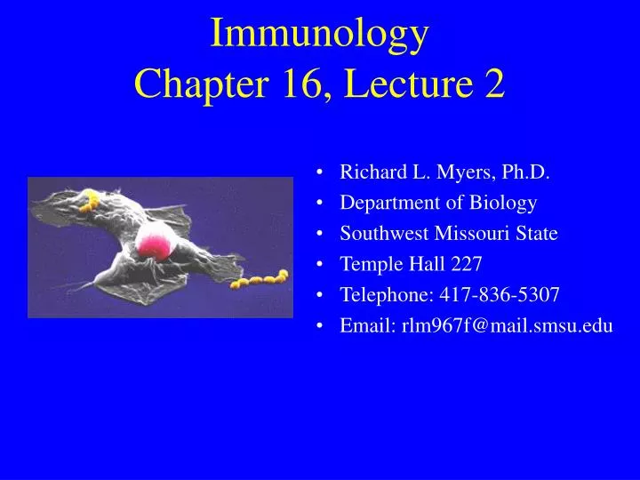
Immunology Chapter 16, Lecture 2. Richard L. Myers, Ph.D. Department of Biology Southwest Missouri State Temple Hall 227 Telephone: 417-836-5307 Email: rlm967f@mail.smsu.edu. The Humoral Response. Used for eliminating extracellular pathogens produces many different antibody molecules

E N D
ImmunologyChapter 16, Lecture 2 • Richard L. Myers, Ph.D. • Department of Biology • Southwest Missouri State • Temple Hall 227 • Telephone: 417-836-5307 • Email: rlm967f@mail.smsu.edu
The Humoral Response • Used for eliminating extracellular pathogens • produces many different antibody molecules • each specific for a certain epitope • may produce 1011 different antibodies • In addition, the constant portion of the antibody may account for biological effector functions
Humoral process requires participation of other cells • macrophages • B cells • also important is the interaction between TH and antigen-class II MHC complex • B cells are the principle cell in humoral immunity • they interact with antigen via a BCR • proceeds with receptor-mediated endocytosis • unlike macrophage which phagocytizes anything • then antigen presented with a class II MHC on the membrane
Humoral effector functions • Activate complement system • Enhance phagocytosis via opsonins • Neutralize bacterial toxins • Neutralize viruses • Prevent colonization at mucosal surfaces • Involved in ADCC
Basic facts • Immunocompetent B cells possess IgM and IgD membrane bound antibodies • Clonal proliferation and differentiation occur after activation • B cells have average cell cycle of 15 hr • Unless activated by antigen, they will die in a few days (usually 90% will die) • Marrow produces about 107 B cells/day
General response to antigen • The response is characterized by the 1) production of antibody-secreting cells and 2) memory B cells • during the lag phase cells undergo clonal selection • then the logarithmic phase occurs • increase in antibody; it eventually declines • for example, with SRBCs, lag phase lasts 4 days; peak plasma cell levels within 5 days; peak antibody within 7 days • IgM secreted initially, followed by IgG • Referred to as the primary response
Primary response with formation of antibodies differs depending upon • nature of the antigen • route of antigen administration • presence of adjuvants • species or strain
Plasma cell • Secondary response different from primary • response is more rapid • produces more antibody • lasts for a longer time • maybe 1,000 times more antibody produced • Secondary response occurs with second exposure to the antigen • Depends upon the existence of memory B cells and memory T cells
Hemolytic plaque assay • Assay to measure plasma cell numbers in mice primed with SRBCs • many modifications • Assay can be used to quantitate plasma cells secreting antibodies specific for any antigen • First, immunize mice with SRBCs
Prepare a spleen cell suspension from a primed mouse • Mix in warm, melted agar to which SRBCs have been added • Prepare a petri dish with a layer of hard agar • Overlay with mixture above • Allow to cool and solidify • Incubate for 1 hr at 37oC
During incubation, antibodies diffuse into agar and binds to the SRBC • Guinea pig serum containing complement is added • Complement reacts with the bound antibody • mediates lysis • Lysis is indicated by a plasma cell surrounded by a clear plaque devoid of cells • Plaques can be counted • referred to as direct plaque-forming cells (PFC)
Elispot assay • Plasma cells quantitated without SRBCs • Use antigen-primed splenocytes • Plate in agar containing antigen • Plasma cells secrete antibody which binds to the antigen • Remove cells • Visualize bound antibody with ELISA
Associative (linked) recognition • This is a process where TH and B cells must see peptides on the same molecule for B cell activation to occur • In the following example the epitope is a viral coat (spike) protein • T cells recognize internal protein which allows B cells to make antibody to coat protein
The activated TH cell recognizes the processed peptide together with the class II MHC molecule • Antibodies can then be produced to the peptide • Binding of antibody to virus occurs • There is also localized release of cytokines • Cytokines allow B cell to proliferate/differentiate
There are other membrane receptors involved • LFA-1 and CD4 are involved in cellular adhesion • Once in contact a signal generates the expression of CD40L on the T cell • This interacts with CD40 on the B cell membrane • This causes induction of cytokine receptors • Results in fully activated B cells • these can proliferate
Assignment • Read Chapter 17, Hypersensitivity Reactions • Review question 3 (pg 439)