Download
1 / 1
10 likes | 120 Vues
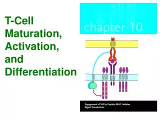
T-cell Phenotypic Profile in Pregnancy: Link Between Immune Activation and Exhaustion N Shah 1 , N Imami 1 , M Johnson 1 . 1 Imperial College London, Chelsea & Westminster Hospital, London, UK. SW10 9NH. Introduction

E N D
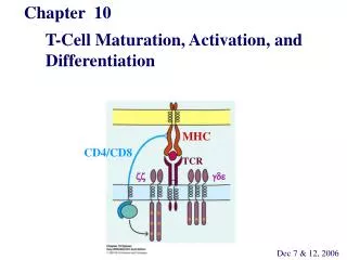
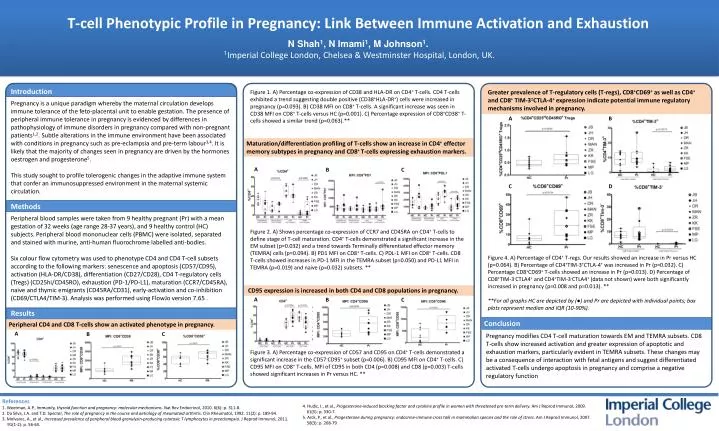
T-cell Phenotypic Profile in Pregnancy: Link Between Immune Activation and Exhaustion N Shah1, N Imami1, M Johnson1.1Imperial College London,Chelsea & Westminster Hospital, London, UK. SW10 9NH Introduction Pregnancy is a unique paradigm whereby the maternal circulation develops immune tolerance of the feto-placental unit to enable gestation. The presence of peripheral immune tolerance in pregnancy is evidenced by differences in pathophysiology of immune disorders in pregnancy compared with non-pregnant patients1,2. Subtle alterations in the immune environment have been associated with conditions in pregnancy such as pre-eclampsia and pre-term labour3,4. It is likely that the majority of changes seen in pregnancy are driven by the hormones oestrogen and progesterone5. This study sought to profile tolerogenic changes in the adaptive immune system that confer an immunosuppressed environment in the maternal systemic circulation. Methods Peripheral blood samples were taken from 9 healthy pregnant (Pr) with a mean gestation of 32 weeks (age range 28-37 years), and 9 healthy control (HC) subjects. Peripheral blood mononuclear cells (PBMC) were isolated, separated and stained with murine, anti-human fluorochrome labelled anti-bodies. Six colour flow cytometry was used to phenotype CD4 and CD4 T-cell subsets according to the following markers: senescence and apoptosis (CD57/CD95), activation (HLA-DR/CD38), differentiation (CD27/CD28), CD4 T-regulatory cells (Tregs) (CD25hi/CD45RO), exhaustion (PD-1/PD-L1), maturation (CCR7/CD45RA), naive and thymic emigrants (CD45RA/CD31), early-activation and co-inhibition (CD69/CTLA4/TIM-3). Analysis was performed using FlowJo version 7.65 . Results Greater prevalence of T-regulatory cells (T-regs), CD8+CD69+as well as CD4+ and CD8+ TIM-3±CTLA-4+ expression indicate potential immune regulatory mechanisms involved in pregnancy. Figure 1. A) Percentage co-expression of CD38 and HLA-DRon CD4+ T-cells. CD4 T-cells exhibited a trend suggesting double positive (CD38+HLA-DR+) cells were increased in pregnancy (p=0.093). B) CD38 MFIon CD8+ T-cells. A significant increase was seen in CD38 MFI on CD8+T-cells versus HC (p=0.001). C) Percentage expression of CD8+CD38+ T-cells showed a similar trend (p=0.063).** A B Maturation/differentiation profiling of T-cells show an increase in CD4+ effector memory subtypes in pregnancy and CD8+ T-cells expressing exhaustion markers. A B C C D Figure 2. A) Shows percentage co-expression of CCR7 and CD45RA on CD4+ T-cells to define stage of T-cell maturation. CD4+ T-cells demonstrated a significant increase in the EM subset (p=0.032) and a trend towards Terminally differentiated effector memory (TEMRA) cells (p=0.094). B) PD1 MFI on CD8+ T-cells. C) PDL-1 MFIon CD8+ T-cells. CD8 T-cells showed increases in PD-1 MFI in the TEMRA subset (p=0.050) and PD-L1 MFI in TEMRA (p=0.019) and naive (p=0.032) subsets. ** Figure 4. A) Percentage of CD4+ T-regs. Our results showed an increase in Pr versus HC (p=0.064). B) Percentage of CD4+TIM-3+CTLA-4+ was increased in Pr (p=0.032). C) Percentage CD8+CD69+ T-cellsshowed an increase in Pr (p=0.013). D) Percentage of CD8+TIM-3-CTLA4+ and CD4+TIM-3-CTLA4+ (data not shown) were both significantly increased in pregnancy (p=0.008 and p=0.013). ** **For all graphs HC are depicted by (●) and Pr are depicted with individual points; box plots represent median and IQR (10-90%). CD95 expression is increased in both CD4 and CD8 populations in pregnancy. A B C Conclusion Peripheral CD4 and CD8 T-cells show an activated phenotype in pregnancy. A B C Pregnancy modifies CD4 T-cell maturation towards EM and TEMRA subsets. CD8 T-cells show increased activation and greater expression of apoptotic and exhaustion markers, particularly evident in TEMRA subsets. These changes may be a consequence of interaction with fetal antigens and suggest differentiated activated T-cells undergo apoptosis in pregnancy and comprise a negative regulatory function Figure 3. A) Percentage co-expression of CD57 and CD95 on CD4+ T-cells demonstrated a significant increase in the CD57-CD95+ subset (p=0.006). B) CD95 MFI on CD4+ T-cells. C) CD95 MFIon CD8+ T-cells. MFI of CD95 in both CD4 (p=0.008) and CD8 (p=0.003) T-cells showed significant increases in Pr versus HC. **