Download
1 / 11
110 likes | 201 Vues
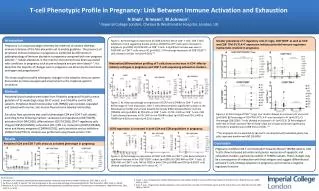
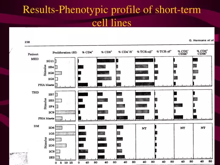
Results-Phenotypic profile of short-term cell lines. Conclusion-Figure 3. CD8 + T cells were stimulated in 2 out of the 3 patients Phenotype of responding cells differed depending on the vaccine clones used Overall cytokine response was seen from TCR- ab +

E N D
Conclusion-Figure 3. • CD8+ T cells were stimulated in 2 out of the 3 patients • Phenotype of responding cells differed depending on the vaccine clones used • Overall cytokine response was seen from TCR-ab+ • All the clones stimulated Natural Killer cells
Rationale-Figure 4. • To determine the type of immune response that is generated by T cell vaccination • Non-stimulated and stimulated PBMC cultures were studied all in vitro
Conclusion-Figure 4. • Controls PBMC cultures did not produce IL-4 • Some control cases showed low levels of IFN-g • IFN-g and TNF-a production was observed in most cultures • IFN-g production increased in most patients after the 3rd vaccination
Rationale-Figure 5. • To study the relative contributions of CD4+, CD8+, and gd T cells to cytokine production after vaccination • Certain T cells were removed from the PBMC • Controls were PBMC containing all T cells • Depleted cell mixtures were irradiated in vitro with vaccine cells • Results were obtained using flowcytometry
Figure 5.-Proliferation and cytokine production of depleted PBMC
Conclusion-Figure 5. • CD4 cells most often reduced proliferation • Depletion of CD8+ T cells did not greatly alter proliferation • In some cases, gd T cell depletion raised proliferative activity • Depletion of gd T cells did not have a major effect on cytokine production • The major effector of cytokine production was CD4+ T cells
Conclusion • Precise mechanisms by which T cell vaccination ameliorates autoimmune disease are still unclear • The majority of patients the proliferative response was maximal after the 2nd and 3rd vaccinations • For long term T cell response, SI units greater than 2 were observed in at least one clones for all patients (See Figure 2) indicating there may be memory antigen-specific lymphocytes
Conclusion Cont’d • From Figure 3., it is concluded that the phenotypic characteristics obtained after stimulation with vaccine cell lines demonstrate that CD4+/- and CD8+/- cells respond to the vaccine • Figure 4., reinerates the fact that cytokine production increased mainly after the 3rd vaccination • Depletion of CD4+ T cells led to a substanial reduction in proliferative response indictaing CD4+ T cells role to vaccine responses in vitro
References • Hermans, Guy, Ulrike Denzer, Ansgar Lohse, Jef Raus, and Piet Stinissen. 1999. Cellular and Humoral Immune Responses Against Autoreactive T cells in Multiple Sclerosis Patients After T cell Vaccination. Journal of Autoimmunity 13: 233-246. • Nester, Eugene W., C. Evans Roberts, Nancy N. Pearsall, Denise G. Anderson, and Martha T. Nester. 1998. Microbiology: A Human Perspective. 2nd edition. Boston: WCB McGraw-Hill. 367-370pp. • Pouly, Sandrine and Jack P. Antel. 1999. Multiple Sclerosis and Central Nervous System Demyelination. Journal of Autoimmunity 13:297-306. • Steinman, Lawrence. 1996. Multiple Sclerosis: A Coordinated Immunological Attack against Myelin in the Central Nervous System. Nature 365: 642-644. • http://mail.med.upenn.edu/~hessd/Lesson2.html