Download
1 / 30
310 likes | 342 Vues
Development of the Heart. 212 – 2004 – Week 6 Avinash Bharadwaj. Retrospect…. Trilaminar embryo Three germ layers Head and tail folds. Cardiovascular System. The Heart Blood vessels Abnormal development Correlation. Some Special Features. Early development and function

E N D
Development of the Heart 212 – 2004 – Week 6 Avinash Bharadwaj
Retrospect… • Trilaminar embryo • Three germ layers • Head and tail folds
Cardiovascular System • The Heart • Blood vessels • Abnormal development • Correlation
Some Special Features • Early development and function • Requirements of the foetus • Non-functional lungs • Placental circulation • Planning for postnatal life • Right-left passage essential during foetal life, obliteration required immediately after birth. • The phylogenetic sequence.
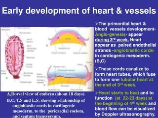
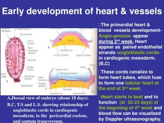
Earliest Development • Angiogenic cell clusters or Blood Islands • Blood cells and endothelium
Cardiogenic Area • Flat embryo • Position of cardiogenic area
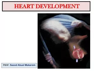
Head Fold • Cardiac area • Gut formation • Pericardium and heart tube
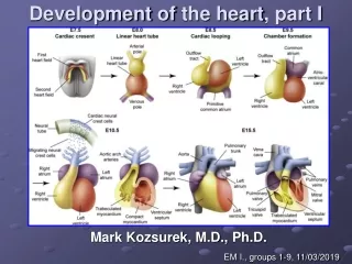
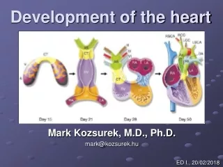
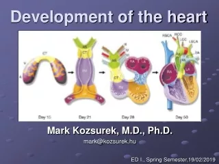
The Heart Tube • Venous and arterial ends • Regions defined • Bulbus cordis • Ventricle • Atrium • Sinus venosus • Conus and Truncus…?
B V D A V SV The Tube Bends
The Chambers Left Front A A A BVL V
A-Ar LA RA AVC The Interior • R and L atria • Atrioventricular canal : common • Outflow • Aortic arches
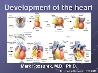
Left – Right Partitioning • Interatrial septum • Interventricular septum • Spiral (aortico-pulomonary septum • Endocardial cushions (A-V cushions) • Functional requirements • There must always be a right to left passage!
Interatrial septum • Partitioning • Right to left passage • Mechanism for closing the passage
A V
Septum Primum • This is a sagittal section seen from the right. AVC V
Foramen Primum • Foramen primum : Between the septum and the AV Cushions
Passage is a Must! • Foramen secundum • Foramen primum about to disappear
Septum Secundum • To the right of primum • Foramen primum has disappeared
Foramen ovale • F. Ovale – • In septum secundum • Further…
The ‘Valve’ • Two septa • Two foramina
Sinus Venosus • Originally a symmetrical structure • Right and left “horns” • Venous return more to the right • Left horn becomes smaller • Opening shifts to the right • Later – part of right atrium
Left Atrium • Four pulmonary veins • Common opening • “Absorption” of veins into atrium • Rough part - auricle
The Ventricular Septum Three Parts • Interventricular septum • AV Cushions • Spiral Septum
The Ventricular Septum R Membranous Spiral (Aorticopulmonary) Muscular
Foetal Circulation • Very little pulmonary flow • Placental Circulation • Right to Left Passages
IVC : Blood from placenta • Ductus venosus • F. ovale • Ductus arteriosus
Changes At Birth • Closure of interatrial septum • Closure of ductus arteriosus • Closure of ductus venosus
Congenital Heart Disease • Septal Defects – Atrial and Ventricular • Endocardial cushion defects • Aorticopulmonary defects • PDA • Others