Download
1 / 35
550 likes | 1.2k Vues
Heart Development. Dr. Nimir. Objectives: Understand early development of blood vessels. B asic understanding of the early stages of heart development. Describe the formation and position of the heart tube. Discus the development of sinus venosus .

E N D
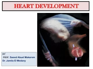
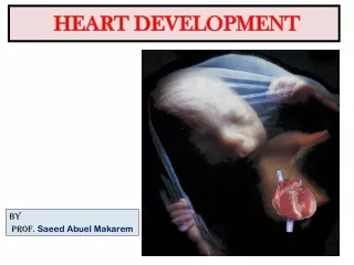
Heart Development Dr. Nimir
Objectives: Understand early development of blood vessels. Basic understanding of the early stages of heart development. Describe the formation and position of the heart tube. Discus the development of sinus venosus. Describe partioning by septa and chambers formation. Discus congenital malformations. Gain knowledge of fetal circulation.
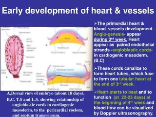
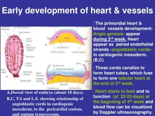
1. Development of Early Blood Vessels 1.1 Yolk sac → blood islands→ endothelia →vessels primitive blood cells.
1.2 Chorion, body stalk, embryonic body → blood vessels →3 separate circulations: vitelline, chorionic, and intraembryonic.
2. Development of Primitive Heart Tube • 2.1 Primordium: Cardiogenicarea→ • Intraembryonic coelom→pericardial coelom; • 2 lateral cardiogenic plates →endocardialheart tubes.
2.2 Primitive heart tube 1) Lateral fold: 2 heart tubes → singleheart tube.
2) Head fold:pericardial coelom → ventral to heart tube • ↘caudal to oropharyngeal membrane
3) Wall of primitive heart tube • Endocardial heart tube→ endocardium • Myoepicardial mentle→ myocardium, epicardium • Cardiac jelly→ subendocardial tissue
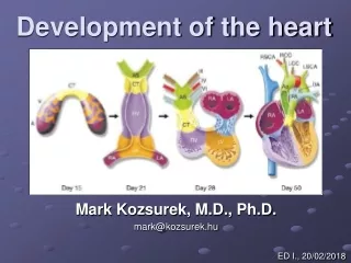
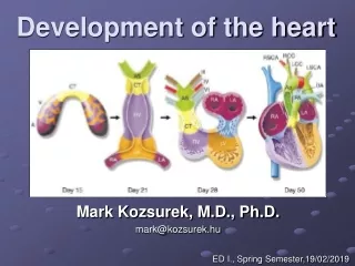
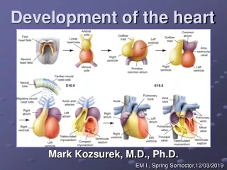
3. Formation of Heart Loop 3.1 Heart tube→bulbus cordis, ventricle, atrium→truncus arteriosus,conus cordis, ventricle, atrium, sinus venosus
3.2 Bulboventricular portion→bulboventricular loop→ cephalic portion bends ventrally,caudally and slightly to the right 3.3 Atrium→ dorsocranially and bulges laterally on each side of bulbus 3.4 Proximal part of bulbus → primitive right ventricle
4. Partitioning of Heart Chambers 4.1 Division of atrioventricular canal Subendocardial tissue→ 2 endocardial cushions→ fuse → right and left canals
4.2 Partitioning of primitive atrium 1) Septum primum→ endocardial cushions →foramen primum.
2) Septum primumabsorbed → foramen secundum→ foramen primum closing 3) Septum secundum→ cover the foramen secundum → foramen ovale
4) Blood from right to left atrium foramen ovale
4.3 Development of sinus venosus • Right horn enlarges (due to left-to-right shunts of blood in venous system) • Sinus-atrial orifice → right; • Receives sup. and inf. vena cava; • Right horn → right atrium (smooth walled part).
2) Left horn degenerates → coronary sinus, oblique vein of left atrium; • Pulmonary vein and its branches → left atrium (smooth walled part)
4.4 Partitioning of the primitive ventricle 1) Apical ventricle wall →muscular interventricular septum→ interventricular foramen
2) Endocardial cushion, right and left bulbar ridges→ membranous interventricularseptum→ interventricular foramen closed
4.5 Division of truncus and bulbus • Truncal ridges +Bulbar ridges→ • aorticopulmonary septum
2)Aorticopulmonary septum→ spiral course → • pulmonary trunk→ right ventricle • aorta → left ventricle
3) Truncus swellings → hollowed out at upper surface → semilunar valves
5. Circulation before and after Birth 5.1 Circulation before birth • Placental circulation: umbilical A. & V. • ductus venosus • foramen ovale • ductus arteriosus
5.2 Changes after birth • Umbilical arteries→ lateral umbilical ligaments • Umbilical vein→ ligamentum teres hepatis • Ductus venosus→ venous ligament • Ductus arteriosus→ arterial ligament • Foramen ovale → oval fossa
6. Congenital Malformations • 6.1 Atrial septal defect • Excessive resorption of the septum primum; • Inadequate development of the septum secundum.
6.2 Ventricular septal defect • Defect of the membranous septum, isolated or associated with other abnormalities.
6.3 Tetralogy of Fallot • Unequal division of conus cordis; • 4 defects: pulmonary stenosis,overriding aorta,ventricular septal defect,hypertrophy of right ventricle; • Causing cyanosis.
6.4Persistent truncus arteriosus • Truncoconal ridges fail to fuse and descend; • Truncus overrides both ventricles; • Accompanied by ventricular septal defect; • Cyanosis, blood to lungs increased.
6.5 Transposition of great vessels • Truncoconal septum failing to follow its spiral course and descending straight downward; • Aorta originates from right ventricle, pulmonary artery from left; • Usually combined with patent ductus arteriosus.
6.6 Patent ductus arteriosus • Ductus arteriosus fails to be closed after birth; • Isolated or combined with other defects.
6.7 Tricuspid atresia • Absence or fusion of tricuspid valves; • Patent oval foramen & ventricular septal defect; • Underdeveloped right ventricle
6.8Pulmonary valvular atresia (or stenosis) • Pulmonary valves are fused for variable distance; • Hypoplastic right heart; • Patent oval foramen and patent ductus arteriosus.
6.9Aortic valvular atresia and stenosis • Aortic valves are fused for variable distance; • Aorta, left ventricle, left atrium underdeveloped; • Accompanied by patent ductus arteriosus.