Download
1 / 15
150 likes | 257 Vues
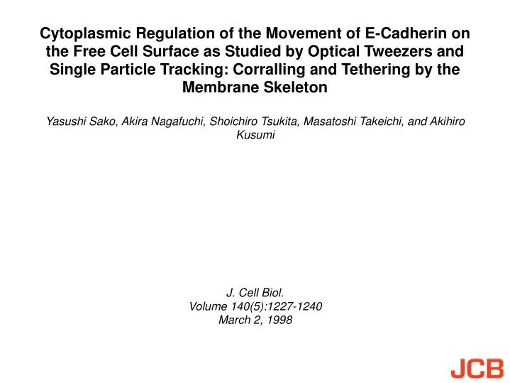
Cytoplasmic Regulation of the Movement of E-Cadherin on the Free Cell Surface as Studied by Optical Tweezers and Single Particle Tracking: Corralling and Tethering by the Membrane Skeleton. Yasushi Sako, Akira Nagafuchi, Shoichiro Tsukita, Masatoshi Takeichi, and Akihiro Kusumi. J. Cell Biol.

E N D
Cytoplasmic Regulation of the Movement of E-Cadherin on the Free Cell Surface as Studied by Optical Tweezers and Single Particle Tracking: Corralling and Tethering by the Membrane Skeleton Yasushi Sako, Akira Nagafuchi, Shoichiro Tsukita, Masatoshi Takeichi, and Akihiro Kusumi J. Cell Biol. Volume 140(5):1227-1240 March 2, 1998
A tether model (A) and a fence model (B) proposed as mechanisms for the regulation of lateral movements of E-cadherin (Sako and Kusumi, 1995) Sako Y. et.al. J. Cell Biol. 1998:140:1227-1240
Structures of the wild-type E-cadherin and its artificial mutants used in this study Sako Y. et.al. J. Cell Biol. 1998:140:1227-1240
Indirect immunofluorescence staining of E-cadherin on the surface of cells transfected with E-cadherin cDNAs Sako Y. et.al. J. Cell Biol. 1998:140:1227-1240
Trajectories of particle–cadherin complexes dragged by OT Sako Y. et.al. J. Cell Biol. 1998:140:1227-1240
(A) The time course of a representative dragging experiment Sako Y. et.al. J. Cell Biol. 1998:140:1227-1240
Distributions of the escape distances Sako Y. et.al. J. Cell Biol. 1998:140:1227-1240
Distributions of the freely dragged distances Sako Y. et.al. J. Cell Biol. 1998:140:1227-1240
Distributions of the effective spring constant (kmsk) of the portions of the membrane skeleton/cytoskeleton that are involved in corralling or tethering E-cadherin molecules Sako Y. et.al. J. Cell Biol. 1998:140:1227-1240
kmsk (effective spring constant) plotted against the freely dragged distance (dfd) for individual particles Sako Y. et.al. J. Cell Biol. 1998:140:1227-1240
SPT trajectories of E-cadherins recorded for 16.7 s (500 video frames) Sako Y. et.al. J. Cell Biol. 1998:140:1227-1240
Distributions of Dmicro Sako Y. et.al. J. Cell Biol. 1998:140:1227-1240
Classification of trajectories into simple Brownian, confined, and directed movement modes in a time window of 5 s Sako Y. et.al. J. Cell Biol. 1998:140:1227-1240
Typical trajectories of Wild (A) and Fusion (B) classified as directed movement Sako Y. et.al. J. Cell Biol. 1998:140:1227-1240
Models of the mechanisms for the regulation of the movement of E-cadherins in the plasma membrane Sako Y. et.al. J. Cell Biol. 1998:140:1227-1240