Download
1 / 20
210 likes | 354 Vues
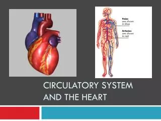
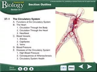
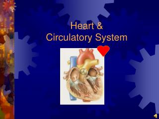
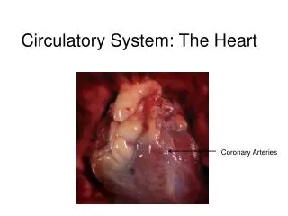
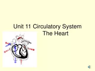
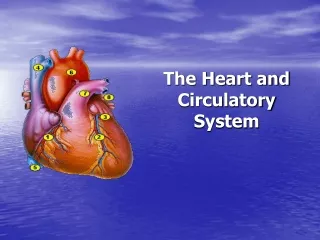
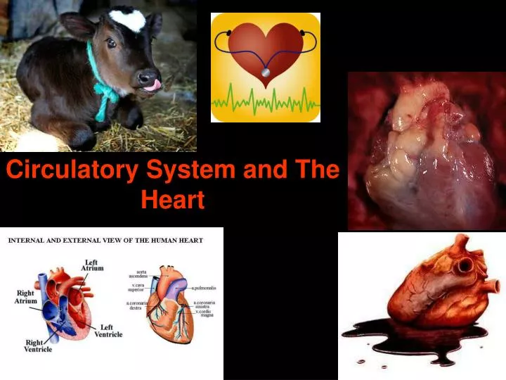
Circulatory System and The Heart. The Heart. Size of fist - 300g Beats 70 times per minute Not a single pump, but two parallel pumps separated by a large muscle called a septum. Pulmonary Circulatory System. Vessels that carry blood to and from the lungs. Systematic Circulatory System.

E N D
The Heart • Size of fist - 300g • Beats 70 times per minute • Not a single pump, but two parallel pumps separated by a large muscle called a septum
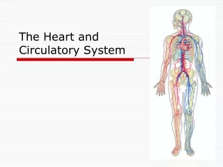
Pulmonary Circulatory System • Vessels that carry blood to and from the lungs
Systematic Circulatory System • Vessels that carry blood to and from the body http://www.nelson.com/ABbio20-30/teacher/protect/otr/Bio2030OTR/attachments/i_AnimationSimulation/blood_circulation.html
Systemic Circulatory System Pulmonary Circulatory System Vessels that carry blood to and from the heart Vessels that carry blood to and from the body
Parts of the Heart – label diagram • aorta • Atrioventricular valve (tricuspid and bicuspid) • Semilunar valves • Septum • Right Atrium • Left Atrium • Right Ventricle • Left Ventricle • Superior vena cava • Inferior vena cava • Pulmonary veins (R/L) • Pulmonary artery (R/L) Be able to describe the path of a blood cell from when it ENTERS the heart from the body to when it LEAVES the heart to the body ***Obstacle course***
Heart Movement • When the ventricles contract, blood is pushed out of the ventricles, through the semi-lunar valves, and into the arteries **Note the AV valves are closed during this time** • When the ventricles relax, the AV valves open, allowing blood to flow into the atria and from the atria into the ventricles **Note the semi-lunar valves are closed at this time**
One Way Blood Flow • Blood circulates through the body in one direction only • AV and Semi-lunar valves inside the heart, prevent the back flow of blood • AV valves are supported by thick connective tissue called chordae tendinae
Setting the Heart Tempo • Cardiac muscle differ from other types of muscles • Cardiac muscle can contract without external nerve simulation; myogenic muscle • Different parts of the heart beat at different tempos individually, but together when united
SA Node • The heart’s tempo is is set by the sinoatrial or SA node • The SA node is a bundle of specialized nerve and muscle cells located where the venae cavae enter the atrium • This bundle sets the hearts tempo at about 70 beats per minute
http://www.nelson.com/ABbio20-30/teacher/protect/otr/Bio2030OTR/attachments/i_AnimationSimulation/cardiac_conduction.htmlhttp://www.nelson.com/ABbio20-30/teacher/protect/otr/Bio2030OTR/attachments/i_AnimationSimulation/cardiac_conduction.html AV Node • The contractions are carried throughout the heart to a second node, the AV node or atrioventicular valve • The AV node conducts the impulse through the septum and towards the ventricles • Thus, the atrium contract prior to the ventricles
Heart Sounds • The heart makes a two-beat sound - “lubb - “dubb” • The sound is created when pairs of valves close • The lubb sound is louder, and is created when the AV valves close, semi-lunar Valves open • The dubb sound is softer, and is created when the semi-lunar valves close, AV valves open
Diastolic • Used to describe the relaxation of the heart • Blood moves through the atria into the ventricles during relaxation
Systolic • Used to describe the contraction of the heart • blood is pushed out of the ventricles upon contraction
Activity – in Text • Page 311 – Exploration
Arteries and Arterioles • Carry blood AWAY from the heart • Where you feel your pulse (change diameter when the heart pumps blood through them) • Thick, muscular walls can withstand high pressure • Blood pumped from heart maintains one way blood flow • Carry oxygenated blood EXCEPT pulmonary artery
Veins and Venules • Carry blood TO the heart • Thin walls, cannot withstand much pressure • Valves maintain one way flow • Skeletal muscles contract – increase pressure – vein opens • Carry deoxygenated blood EXCEPT for pulmonary vein • When the muscle relaxes, the vein widens, forcing blood to build up at the valves • When skeletal muscles contract, the vein narrows, and the valves open, forcing blood to pass through to the heart **Store about 65% of your blood **
Capillaries • Thinnest blood vessel (SIZE: one blood cell thick) • Very weak - bruising • Where venules and arterioles connect • Site of fluid and gas exchange