Download
1 / 26
270 likes | 477 Vues
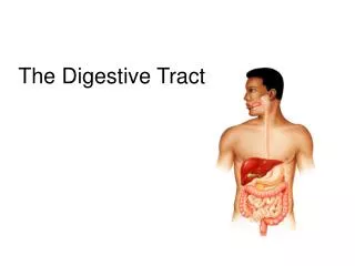
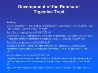
Lab 9. Digestive Tract (2) &Digestive glands. Small intestine (N0.1) Large intestine (No.26) Pancreas (No.29) Liver (No.30) Exercise. 20131127. Small intestine. Mucosa epi. lamina propria muscularis mucosa Submucosa Muscularis externa Serosa. Luminal surface modification:

E N D
Lab 9. Digestive Tract (2) &Digestive glands Small intestine (N0.1) Large intestine (No.26) Pancreas (No.29) Liver (No.30) Exercise 20131127
Small intestine • Mucosa epi. lamina propria muscularis mucosa • Submucosa • Muscularis externa • Serosa Luminal surface modification: 1.Plicae circulares 2.Intestinal villi 3.Microvilli
Mucosa • Submucosa • Muscularis externa • Serosa Human jejunum
Villus • Epithelium 1.absorptive cell striated border (microvilli) 2.Goblet cell • Lamina propria CT+SM
Intestinal glands • Columnar cells • Goblet cells • Paneth cells • at the base of the crypts • 3-5 in a group • Acidophilic G.
Muscularis externa • Myenteric plexus
Serosa • CT • Mesothelium
Large intestine Small intestine
No villi • No Paneth cell • Numerous Goblet cells
Pancreas • Capsule • Septa extend into the gland • Ill-defined lobule
Exocrine gland Endocrine gland
Exocrine portion: pyramidal serous cells centroacinar cells
Intercalated ducts • Intralobular ducts • Interlobular ducts
Interlobular ducts • Islets of Langerhands: scattered among the exocrine portion
Liver • Capsule • Parenchyma: hepatocyte
Liver lobule • Central vein • Plates of hepatocytes • Sinusoids
Liver plates • Liver sinusoids: kupffer cells
Portal area: Interlobular veins Interlobular arteries Interlobular bile ducts
Human liver Pig liver