Download
1 / 55
650 likes | 1.13k Vues
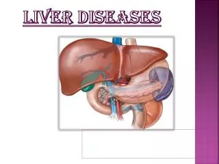
Liver diseases. Review Outline. Hepatology Abnormal LFTs Viral hepatitis (A, B, C) PSC PBC AIH NASH/NAFLD Drugs (Tylenol) Cirrhosis Ascites Metabolic liver diseases (A1AT, HH, Wilsons) Gilbert’s Disease Liver masses Pregnancy Pancreatiobiliary Pancreatitis – acute and chronic

E N D
Review Outline • Hepatology • Abnormal LFTs • Viral hepatitis (A, B, C) • PSC • PBC • AIH • NASH/NAFLD • Drugs (Tylenol) • Cirrhosis • Ascites • Metabolic liver diseases (A1AT, HH, Wilsons) • Gilbert’s Disease • Liver masses • Pregnancy • Pancreatiobiliary • Pancreatitis – acute and chronic • Pancreatitis complications • Pancreatic cancer • Billiary disease
Hepatology Pearls • Hepatitis: AST and ALT • Cholestasis: TB and ALP • ALT more specific than AST • Measures of function: ALB, Coags, Bili • Alcoholic hepatitis AST>ALT 2-3:1 (NASH with cirrhosis also)
Abnormal LFT’s • Asymptomatic elevation of ALT is most common problem • If isolated and less than 3-fold elevation then stop alcohol or drug and recheck in 2-3 months • If persistent then further workup is needed
Abnormal LFT’s • ALT >10 fold (>400) • Acute viral • Drug/toxin • Ischemic/Budd Chiari • Autoimmune hepatitis • Wilson’s disease
Abnormal LFT’s • Modest ALT (<300) has a wide differential • Usually EtOH or chronic viral hepatitis • Remember AST:ALT > 2:1 highly suggestive of EtOH • AIH, NASH/NAFLD, Wilson’s, Hemochromatosis, infiltrative/granulomatous dz
Abnormal LFT’s • Mildly high ALP or TB without evidence of biliary dz, think infiltrative (TB, sarcoid, fungal) or metastatic disease • Workup mainly by history and risk factors • Image or biopsy for diagnostic purposes is not always needed
Abnormal ALP • Hepatic • PBC (middle aged women) • PSC (IBD history) • Gallbladder/stone disease • Meds (tetracyclines, OCP’s, ceftriaxone) • Infiltrative liver dz (sarcoid, TB, CA) • Pregnancy • Bone (Mets or Paget’s disease)
Hepatitis A • Fecal-oral transmission • Symptoms: Adult > children • Transplacental transmission occurs • No carrier states, rarely fulminant • Can have cholestasis for up to 6 mos • Vaccine: Patients with liver dz/risks/ travelers • Acute infection: + IgM anti-HAV, Vaccination: + IgG anti-HAV • IG prophylactic for Hep A
Hepatitis B • Incubation 1-6 months • Transmitted sexually, parenterally, mucous membrane exposure • Can present with serum sickness (fever, arthritis, urticaria, angioedema) • Associated with polyarteritis nodosa (PAN)
Hepatitis B Serology • HBsAg - first marker present in pts with active Hep B infection • HBcAg - core inner shell of virion, not seen in serum, does not circulate • HBeAg - soluble protein secreted from hepatocytes, correlates with with both quantity of intact virus, infectivity and liver inflammation
Hep B Serology • Anti-HBc: first Ab to appear, 1°IgM then IgG, persists for life, best marker for previous exposure • Anti-HBe: appears several wks post illness • Anti-HBs: indicates previous exposure to Hep B or the vaccine
HBV Window Period • HBsAg is not detectable • Anti-HBs not yet present • Anti-HBc appears • May be several weeks • Check Anti-HBc IgM to confirm acute infection, otherwise looks like previous exposure
HBV Scenarios Acute infection Carrier Vaccinated Exposed Immune Acute Window Exposed Ab lost
Course of Hep B • 90% self limited, 1-2% fulminant • 5-7% chronic carrier states • Asymptomatic • chronic persistent hepatitis • chronic hepatitis B - risk of hepatocellular CA, cirrhosis • Inverse relationship between age and development of carrier state, 95% of infants-chronic • 5-10% transplacental transmission
HepB vaccine/prophylaxis • 95% of immunocompetent pts develop antibody (anti-HBs) • Only 50% of HD pts develop antibody • May be given to pregnant pts • 3 doses at 1, 2 and 6 months • HBIG Alone: • sexual contacts of carriers and household members of acute Hep B • HBIG + vaccine (exposed is HBsAg negative) • blood exposure to pt w/acute Hep B • newborns of Hep B mothers
Treatment of Chronic Hep B • Interferon: • 4-6 months • 30% long term remission • Lamivudine: • 6 months • Decreases HBV-DNA (does not eradicate) • Reduces ALT levels • 15-20% seroconvert • HBeAg + to - with new anti-HBe • Adefovir (Hepsera)
Hepatitis C • Most common liver disease in the US • IVDU, cocaine use, prisons, blood products prior to 1990, tattoo • Genotype 1 most common in the US • 70-80% of Hep C infected become chronic • 25% carriers • 50% abnl LFT’s but asymp • 25% with chronic active/sx • 25% cirrhosis post 20-25 years of infection • 5% sexual transmission over 10-20 yrs • <5% transplacental transmission
Serological Tests • Third generation anti-HCV+ >95% sensitive • If high pre-test probability and anti-HCV negative can do PCR testing (more often in renal failure or transplant) • Genotype testing required for treatment candidates only
Hepatitis C Therapy • Interferon Alone < 15% • Interferon/Ribavirin • 12 months Genotype 1 • 6 months Genotype 2 and 3 • Sustained response (Hep C PCR- 6 mos post therapy) • 5-15% monotherapy • 35-40% combo therapy • 45-70% PEG combo therapy • Must use birth control--teratogenic
Contraindications for Therapy • HB<12 female, <13 male • WBC<1500 • Plt<100,000 • Severe psych dz (depression) • Pregnancy • Decompensated Cirrhosis • CAD • Autoimmune diseases
Extrahepatic Manifestations • Glomerulonephritis/MPGN • Cryoglobulins • Porphyria cutanea tarda (PCT) • Thrombocytopenia • Autoantibody • ITP • Neuropathy • Thyroiditis • Sjogren’s Syndrome • Inflammatory arthritis
Hepatitis D • Requires coexistent B • Usually found in IVDA • Coinfection: does not worsen acute Hep B or risk for chronic state • Superinfection: frequently severe/fulminant • Dx: Anti-HDV IgM
Hepatitis E • Monsoon flooding • Fecal-oral route • No chronic forms • Fulminant hepatitis in 3rd trimester of pregnancy
Chronic Hepatitis • By definition, active hepatitis for 6 months or longer • Usually viral but also metabolic and autoimmune
Case 38 yo woman presents to your office c/o fatigue and pruritis for 6 months. Routine bloodwork reveals normal CBC, TB, AST and ALT. ALP is elevated at 260. Skin exam shows this:
Primary Biliary Cirrhosis (PBC) • Middle-aged woman • Elevated alkaline phosphatase • Symptom: itching! • Later: jaundice, osteomalacia, osteoporosis • Xanthomas/xanthalasmas • hypercholesterolemia with low risk CAD • Diagnosis: • antimitochondrial antibody (AMA) • liver bx with granulomas • Tx: Ursodeoxycholic acid (Actigall) improves survival; late-liver transplant
Case • 38 yo gentleman with h/o chronic diarrhea and occasional BRBPR who presents to your office with new onset jaundice and fever. • ERCP shows:
Primary Sclerosing Cholangitis (PSC) • Male: female 2:1 • Intra and Extrahepatic inflammation and sclerosis of the biliary tree • Strong Association (75%) with IBD (Ulcerative colitis) • Cholestatic enzyme pattern (TB and ALP) • Dx: ERCP or cholangiogram • ~15% develop cholangiocarcinoma • Tx: Stenting of dominant strictures, Liver Transplant
Autoimmune Hepatitis • Young women • insidious hepatitis, elevated AST/ALT • assoc with other autoimmune dz • hypothyroid, ITP, Coombs+ anemia • Dx: • Serology: ANA+, Anti-smooth muscle Ab+ (most specific), Elevated quant IgG, Anti-LKM • Histology: Plasma cells, interface hepatitis • Tx: Prednisone-rapidly reverses sx and improves survival, add azathioprine for steroid sparing
NASH/NAFLD • Risk Factors: • Obesity or gastric bypass • DM • Hyperlipidemia • TPN, amiodarone • Histology: Steatosis, PMN’s, Necrosis • Tx: Wt Loss (>15%), Exercise, Control of DM, Lipid Lowering Agents
Methyldopa Acetaminophen Trazodone Nitrofurantoin Phenytoin INH-1% severe hepatitis, correlates with age OCP’s-cholestasis, benign adenomas, FNH Erythromycin Valproic Acid Methotrexate-indolent cirrhosis Drugs and chronic hepatitis
Acetaminophen • Pt who drinks EtOH and ingests an “over-the-counter” pain medicine develops fulminant hepatitis (AST/ALT/PT) • Toxicity secondary to the depletion of glutathione, which reduces the metabolites. • Malnutrition and EtOH deplete glutathione and therefore lead to toxicity of Tylenol-even therapeutic doses • Tx: N-acetylcysteine • Beware of the “normal” acetaminophen level
Fulminant Hepatic Failure • Acetaminophen, viral, AIH, Wilson’s disease, Budd-Chiari syndrome • Tylenol survival >50%, viral 30-50% • Remember Kings College Criteria • Mainly encephalopathy (III or IV) • pH < 7.3, PT > 100, Cr > 3.4 • Main cause of death: cerebral edema, sepsis • Transfer these patients to a transplant center
Hepatic Masses • HCC > 75% in setting of cirrhosis • Esp. chronic Hepatitis B, Hep C, hemochromatosis, EtOH • Aflatoxin (raw peanuts, raw peanut butter) • alpha-fetoprotein (>100), tender hepatomegaly • Adenomas: on OCP’s. Need resection because of bleeding risk and progression to cancer • Hemangiomas rarely need resection
Cirrhosis • EtOH - most common cause in the U.S. • Esophageal Varices • 1/3 bleed; 1/3 die with bleed • Larger, distal varices bleed • Propranolol rebleeds and may prevent the first bleed • Tx: • Octreotide • Banding or Sclerotherapy • Transjugular intrahepatic portosystemic shunt (TIPS)
Cirrhosis • Encephalopathy • Hyperreflexia and Asterixis • Usually precipitated by infection, bleed, drugs • Tx: Lactulose (acidifies the colonic contents) • Hepatorenal Syndrome • Urinary Na+ < 10, no urine protein • Oliguric Renal Failure • Usually iatrogenic with diuretics/Abx-esp Aminoglycosides
Ascites • Cause: requires peritoneal tap-send for cell count, protein, albumin, cytology • SAAG: Serum albumin-ascitic albumin • >1.1 Portal Hypertension • Budd-Chiari, RHF, Cirrhosis • <1.1 Non Portal Hypertensive causes • TB, Nephrotic syndrome, Pancreatitis and Peritoneal carcinomatosis • Chylous Ascites (lymph blockage) • Trauma, Lymphoma, TB and Filiarisis
Ascites • If protein < 1.0 in cirrhotic ascites then patient is at increased risk for SBP • SBP: • Cloudy fluid, some with fever, abd pain • > 250 PMN’s or >400 total WBC’s • E. coli, Strep, Klebsiella • Treat with cefotaxime • Albumin (1.5 g/kg, then1.0 g/kg on day 3) • Tx: Na+( 2 gm) and water restriction (2 L) • Spironolactone + loop diuretic • Refractory: TIPS
Gilbert’s Syndrome • Benign, chronic disorder glucuronyl transferase • Autosomal dominant with variable penetrance • 7% of the population • Mild Unconjugated hyperbilirubinemia (indirect bilirubin), <5.0 TB. • Bili induced by exercise, EtOH, surgery, infection and fasting • No treatment needed
Hereditary Bilirubin Disorders • Crigler-Najjar Syndrome • moderate to severe unconjugated hyperbilirubinemia • Usually presents in childhood • Dubin-Johnson Syndrome • Low level conjugated hyperbilirubinemia • Pigmented granules in hepatocytes
Question: 49 yo wm with h/o of emphysema presents with elevated AST and ALT. (ALT>AST ) Previous CT Scan of the Chest revealed bulla at bilateral bases. Family history significant for brother with chronic liver disease and social history significant for 1-2 drinks/night. • Suspected Diagnosis for LFT abnormalities?
Alpha-1 Antitrypsin Deficiency • Autosomal recessive • Pt with lung disease and hepatitis • Dx: Alpha1-antitrypsin phenotype • Pi ZZ • Tx: Liver transplant if lung disease does not preclude pt from this. Does not recur in transplanted liver
Hemochromatosis • Autosomal Recessive • Presents in the 50’s with cirrhosis or CHF • Iron deposition in liver, heart, pancreas, and pituitary • Clinical Signs: • Hepatomegaly (95%) • Hyperpigmentation (90%) • Diabetes (65%) • Arthropathy (40%) / CPPD • Cardiac involvement (15%)
Hemochromatosis • 25-30% risk for hepatocellular carcinoma • Diagnosis: • Serum Iron, ferritin (1000’s) and transferrin percent sat > 50%, C282Y homozygotes • Liver biopsy with Iron staining (hepatic iron index) • Treatment: Weekly phlebotomies (Hct 32%), decreased ferritin • prolongs life and improves color/cardiac fx • Men 10x more symptomatic than women