Download
1 / 30
420 likes | 1.34k Vues
CHOLESTASIS & CHOLESTATIC LIVER DISEASES. Dr. Sebati ÖZDEMİR Cerrahpaşa Faculty of Medicine Division of Gastroenterology. DEFINITION. Cholestasis is defined as the failure of normal bile to reach the duodenum.

E N D
CHOLESTASIS& CHOLESTATIC LIVER DISEASES Dr. Sebati ÖZDEMİR Cerrahpaşa Faculty of Medicine Division of Gastroenterology
DEFINITION • Cholestasis is defined as the failure of normal bile to reach the duodenum. • This may be due to pathology anywhere between the hepatocyte and the ampulla of Vater.
DEFINITION • Functionally, cholestasis is defined as a decrease in canalicular bile flow. There is a decreased hepatic secretion of water and/or organic anions (bilirubin, bile acid). • Morphologically, cholestasis is defined as the accumulation of bile in liver cells and biliary passages.
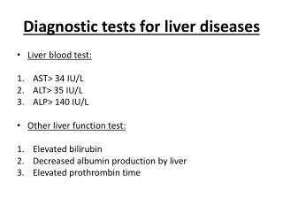
DEFINITION • Clinically,cholestasis is the retention in the blood of all substances normally excreted in the bile. • Serum bile acids, alkaline phosphatase and g-glutamyl-transpeptidase (GGT) are increased. • Clinical features are itching (not always present) and jaundice.
CLASSIFICATION • Cholestasis may be classified as extra- or intra-hepatic, and acute or chronic. • Extra-hepatic cholestasisencompasses conditions where there is physical obstruction to the bile ducts. • Intra-hepatic cholestasisincludes those conditions where there is no demonstrable obstruction (on cholangiography) to the major bile ducts.
EXTRA-HEPATIC CHOLESTASIS • The most common cause is a stone in the common duct. • Other causes are: • carcinoma of the pancreas and ampulla, • benign bile duct stricture, • Cholangiocarcinoma.
INTRA-HEPATIC CHOLESTASIS • Those conditions where there is no demonstrable obstruction (on cholangiography) • to the major bile ducts. • These causes are: • drug-induced cholestasis, • cholestatic hepatitis, • hormones, • Primary biliary cirrhosis • Septisemia • Primary sclerosing cholangitis may causes both intra- and extra-hepatic cholestasis.
CLINICAL FEATURES • Sessiz olabilir: komplikasyon gelişme riski % 1’dir. • Semptomlu olabilir (biliyer kolik): % 2. • Komplikasyonlar: Kolesistit, koledo-kolitiyazis, biliyer obstrüksiyon, akut pankreatit, perforasyon, safra kesesi Ca • En sık görülen:Kolesistit: Ağrı daha uzundur, bulantı, kusma, halsizlik, ateş.
a b (a) MRCP in a 39-year-old woman with right upper quadrant discomfort. Ultrasound showed a bile duct of 1cm but no stones were seen. Gallbladder was normal. Liver function tests were normal apart from marginally abnormal g-GT. The MRCP shows filling defects in the mid bile duct and stones were removed after endoscopic sphincterotomy. (b) MRCP in a patient presenting with cholestasis and change in bowel habit. Both the common bile duct and pancreatic duct are dilated. ERCP showed carcinoma of the ampulla. (c) MRCPin a 40-year-old woman with chronic cholestasis of unknown aetiology. There are dilatations and strictures of the intra-hepatic and peri-hilar bile ducts. Diagnosis: sclerosing cholangitis. c
a ERCP showing: (a) dilated bile duct above a stricture. The pancreatic duct comes to an abrupt halt in the head of the pancreas. Appearances are characteristic of carcinoma of the pancreas; (b) common bile duct filling as far as a hilar stricture due to a cholangiocarcinoma ERCP, normal appearances. C, common bile duct; G, gallbladder; PD, pancreatic duct b
Sphincterotome inserted into ampulla of Vater. The wire has been bowed and the sphincterotomy cut has begun. ERCP showing common bile duct stone. A sphincterotome has been passed into the lower end of the bile duct.
b a c • ERCP showing trawling of bile duct with • balloon catheter to remove stones. • Removal of duct stone with basket. • (c) Naso-biliary tube with stones • in the common bile duct.
(a) ERCP showing malignant stricture (arrows) at lower end of bile duct. (b) Mesh metal stent (Wallstent) placed across the stricture.
Primary Biliary Cirrhosis • Primary biliary cirrhosis (PBC) is a disease of unknowncause in which intra-hepatic bile ducts are progressivelydestroyed. • Circulating antibodies againstmitochondria (AMA) are foundin virtually 100% of patients with PBC
Primary Biliary Cirrhosis • Ninety per cent of patients with PBC are female. Thecause for disease prevalence among women is unknown. • The patient is usually 40–60 years of age. • Widespread use of biochemical screening has resulted inan increasing number of patients being diagnosed whenasymptomatic.
Primary Biliary Cirrhosis • PBC is associated with almost any autoimmune disease. • The collagenoses, especially rheumatoid arthritis, dermatomyositis,mixed connective tissue disease andsystemic lupus erythematosus, are particularly frequent.
Primary sclerosing cholangitis • The condition is of unknown cause. • All parts of thebiliary tree can be involved in a chronic, fibrosing, inflammatoryprocess which results in obliteration of the biliarytree and ultimately in biliary cirrhosis.
Primary sclerosing cholangitis • There is an association with inflammatory bowel disease. There is a 75%association with ulcerative colitis. • The prevalenceof PSC among ulcerative colitis patients is approximately5% with an overall 10–15% prevalance of hepaticabnormalities. • PSC may complicate large bowelCrohn’s disease.