Download
1 / 22
260 likes | 913 Vues
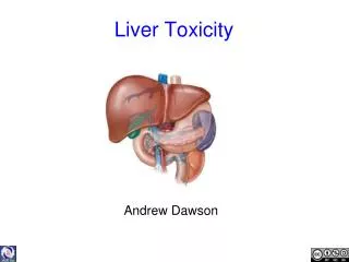
liver. ALI B ALHAILIY. Liver Diagram. Liver Diagram. Liver Function. The liver is one of the largest solid organs of the body. It is located in the upper right part of the abdomen. Most of the organ lies under cover of the rib cage. Liver function

E N D
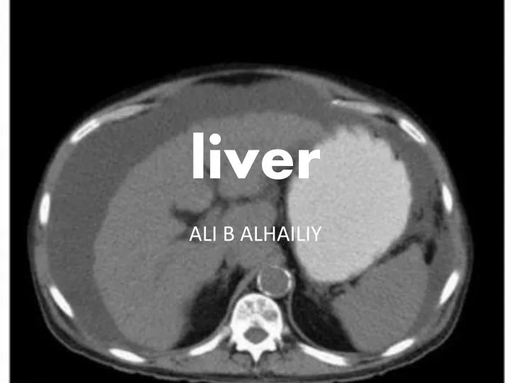
liver ALI B ALHAILIY
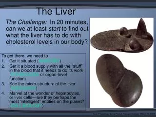
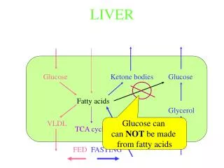
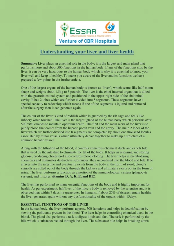
Liver Function • The liver is one of the largest solid organs of the body. It is located in the upper right part of the abdomen. Most of the organ lies under cover of the rib cage. • Liver function • It is not just the liver’s size but also its functions that makes it so important. • Its major functions include processing the food that passes through the gut and converting it into energy that can be utilized by the body. • It is also a powerful detoxification center that handles many chemicals, alcohol, poisons and toxins as well as drugs and clears the blood. • The liver also makes bile and stores it in a small pouch like organ called the gall bladder. This bile helps in digestion especially fats.
Causes of liver disease • Liver diseases may vary in causation. • They may be of short duration, acute liver disease, or long term, chronic liver disease. An acute liver disease may also convert into a chronic liver disease over time. • Some liver diseases are caused by infective viruses like Hepatitis virus(A, B and C). • Liver diseases also result from taking in some drugs or alcohol over long term. Sometimes the diseased liver over long term becomes shrunken and scarred. • Such a condition is called cirrhosis. Like other organs liver can also be affictedwith cancers.
Types of liver disease • Alcohol related liver disease is one of the commonest toxin induced liver disease worldwide. In normal cases the liver breaks down alcohol in the body. • If there is too much intake over a long period of time the liver fails to perform its functions leading to a condition called Alcoholic Liver disease. • The hepatitis phase leads to swelling of the liver and damage. At the initial phases of alcohol liver disease if alcohol is discontinued the changes may be reversed. (3) • Liver disease may also be Non-Alcoholic called Non Alcoholic Fatty Liver Disease. This results in fat deposits on the liver and is seen in obese, diabetic and individuals with high blood cholesterol. • Liver disease is also called hepatic disease. Since it is a large organ, nearly two thirds of the liver has to be affected for the symptoms of hepatic disease to show in most individuals.
If there is very high blood pressure in the portal vein the condition is termed portal hypertension. This can lead to cirrhosis, enlarged abdomen with fluid (ascitis), bleeding, enlarged spleen, and sometimes jaundice. Bleeding may occur in the esophagus or via rectum. • Portal hypertension is often a result of liver cirrhosis that results from chronic liver disease. • Portal hypertension may also sometimes lead to a condition called hepatic encephalopathy where the brain is affected and the person may go into coma. This is usually accompanied by liver failure.
Liver Diagnosis • A- History and physical examination • Diagnosis of liver disease is based on initial history and physical examination. • History of previous illness, drug or alcohol intake, family history of liver disease needs to be evaluated in detail. • History of contaminated blood transfusion, unprotected sexual intercourse or sharing contaminated needles can raise suspicion of a chronic infective liver disease with viruses like Hepatitis B or C.
Liver Diagnosis • B- Laboratory examination • Blood – Initial tests for liver disease include complete blood examination.
Liver Diagnosis • C- Radiological studies • Imaging and radiographical studies are used to detect and confirm liver diseases. These include – • Ultrasonogramof the abdomen – This uses sound waves to detect liver pathology This is useful, inexpensive and a noninvasive method to diagnose several conditions. It is most commonly used to detect gall bladder stones and other pathologies. An USG with Doppler can also look at the blood flow in the portal venous system and diagnose obstructions and portal hypertension. • CT scan – Computed Axial Tomographical image or scan can be used to look at deeper tissues within the liver in detail to diagnose several liver disease conditions. • MRI is a clearer imaging study to look at the damaged liver tissues for clues regarding pathology. • ERCP (Endoscopic retrograde cholangiopancreatography) – In this procedure a long thin tube, called an endoscope, that has a camera at its tip is guided into the gastrointestinal tract to look at the bile and pancreatic ducts. Any pathology in these tracts can be detected. ERCP can be used in diagnosis of tumors, gall bladder stones and obstructions, cysts etc.
Liver Diagnosis • D- Liver Biopsy • This procedure involves using a long thin needle to aspirate bits of tissues of the liver under a local anesthetic agent. The needle is guided into the liver with the help of either USG or CT scan. • This is used to confirm the diagnosis. The aspirated material is examined under the microscope. • A liver biopsy specimen may also be obtained when the patient is operated for. This could be for a liver tumor.
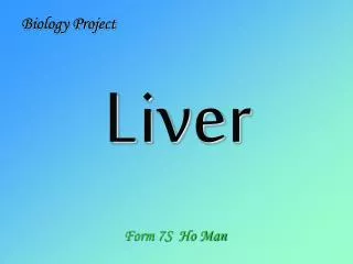
LIVER CANCER (METS) LIVER CYST
LIVER MASS IN CONTRAST ENHANCED T1 W GALL STONE
Liver Cirrhosis & Ascites • Cirrhosis is a result of advanced liver disease. • It is characterized by replacement of liver tissue by fibrosis (scar tissue) • These changes lead to loss of liver function. Cirrhosis is most commonly caused by alcoholism, hepatitis B and hepatitis C, and fatty liver disease, but has many other possible causes.). • Ascites(fluid retention in the abdominal cavity) is the most common complication of cirrhosis. • It is associated with a poor quality of life, increased risk of infection, and a poor long-term outcome. • Cirrhosis is irreversible, and treatment usually focuses on preventing progression and complications. In advanced stages of cirrhosis the only option is a liver transplant
Management • Generally, liver damage from cirrhosis cannot be reversed, but treatment could stop or delay further progression and reduce complications. A healthy diet is encouraged, as cirrhosis may be an energy-consuming process. Close follow-up is often necessary. Antibiotics are prescribed for infections, and various medications can help with itching. Laxatives, such as lactulose, decrease risk of constipation; their role in preventing encephalopathy is limited.
Rupture of the spleen • Rupture of the spleen, an organ in the upper left part of the abdomen, is a situation that requires immediate medical attention. The rupture of a normal spleen can be caused by trauma, such as a motor vehicle accident. • The most common cause of a ruptured spleen is blunt abdominal trauma, such as in traffic or sports accidents. Direct, penetrating injuries, for example, stab or gunshot wounds are rare. • Non-traumatic causes are less common. These include infectious diseases, medical procedures such as colonoscopy, haematological diseases, medications, and pregnancy.
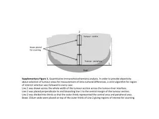
Diagnosis • Traumatic rupture of the spleen on contrast enhanced axial CT (portal venous phase) • Splenic rupture is usually diagnosed by ultrasoundof the abdomen. Ultrasound can detect free fluid due to bleeding and might directly show damage to the spleen itself. • Radiographs of the thorax and abdomen might be performed to exclude other injuries. • In stable patients, CT Scanof the abdomen can give a fuller overview of the splenic and other injuries.