Download
1 / 95
1k likes | 1.3k Vues
GENERAL CONSIDERATIONS ON BONES & JOINTS . 21. September 2011 Wednesday. Kaan Yücel M.D., Ph.D . GENERAL CONSIDERATIONS ON BONES. Osteology ( Gk , osteon, bone, logos, science) is the branch of medicine concerned with the development and diseases of bone tissue. The human skeleton

E N D
GENERAL CONSIDERATIONS ON BONES & JOINTS 21. September2011Wednesday Kaan Yücel M.D., Ph.D.
Osteology (Gk, osteon, bone, logos, science) is the branch of medicine concerned with the development and diseases of bone tissue. The human skeleton 270 bones in the newborn 222 bones in children 206 bones in adults Why Humans Have 270 Bones at Birth, but Only 206 Bones Once We are Adults
Including the hyoid bone (oshyoideum) and the three auditory ossicles (malleus, incus and stapes) on each side, the head (caput) has 29 bones. The vertebral column (columnavertebralis) has 26 bones. Thorax (chest) is composed of 25 bones. The number of the upper limb/extremity bones (ossamembrisuperirois) is 64, whereas that of the lower limb/extremity is 62.
The skeletal system may be divided into • 2functional parts: • The axial skeleton • head (cranium or skull) • neck (hyoid bone and cervical vertebrae) • trunk (ribs, sternum, vertebrae, and sacrum) • The appendicular skeleton • Limbs • including those forming the shoulde & pelvic girdles • 126 bonesin the appendicular skeleton • 80 bonesin the axial skeleton
Bone is a living tissue capable of changing its structure as the result of the stresses to which it is subjected. • Like other connective tissues, bone consists of cells, fibers, and matrix. • It is one of the hardest structures of the animal body, because of the calcification of its extracellular matrix.
Living bones have some elasticity (results from the organic matter) and great rigidity (results from their lamellous structures and tubes of inorganic calcium phosphate). • Its color, in a fresh state, is pinkish-white externally, and deep red within.
Cartilage and Bones • The skeleton is composed of cartilages and bones. • Cartilage is a resilient, semirigid form of connective tissue that forms parts of the skeleton where more flexibility is required—for example, where the costal cartilages attach the ribs to the sternum.
Cartilage and Bones Also, the articulating surfaces (bearing surfaces) of bones participating in a synovial joint are capped with articular cartilage that provides smooth, low-friction, gliding surfaces for free movement.
Cartilage and Bones • Blood vessels do not enter cartilage (i.e., it is avascular); consequently, its cells obtain oxygen and nutrients by diffusion. • The proportion of bone and cartilage in the skeleton changes as the body grows; the younger a person is, the more cartilage he or she has. • The bones of a newborn are soft and flexible because they are mostly composed of cartilage. • Bone has a protective function; the skull and vertebral column, for example, protect the brain and spinal cord from injury.
A fibrous connective tissue covering surrounds each skeletal element like a sleeve, except where articular cartilage occurs; that surrounding bones is periosteum, whereas that around cartilage is perichondrium.
The periosteum and perichondrium nourish the external aspects of the skeletal tissue. • They are capable of laying down more cartilage or bone (particularly during fracture healing) and provide the interface for attachment of tendons and ligaments.
Classification of Bones • Bones are classified according to their shape. • Long bones • Short bones • Flat bones • Irregular bones • Sesamoid bones
Classification of Bones • Long bones are tubular (e.g., the humerus in the arm).
Classification of Bones • Short bones are cuboidal and are found only in the tarsus (ankle) and carpus (wrist).
Classification of Bones • Flat bones usually serve protective functions (e.g., the flat bones of the cranium protect the brain).
Classification of Bones • Irregular bones have various shapes other than long, short, or flat (e.g., bones of the face).
Classification of Bones • Sesamoidbones(e.g., the patella or knee cap) develop in certain tendons and are found where tendons cross the ends of long bones in the limbs; they protect the tendons from excessive wear and often change the angle of the tendons as they pass to their attachments.
There are two types of bones according to histological features: compact bone and spongy (trabecular) bone. • They are distinguished by the relative amount of solid matter and by the number and size of the spaces they contain.
All bones have a superficial thin layer of compact bone around a central mass of spongy bone, except where the latter is replaced by a medullary (marrow) cavity. • Spongy bone is found at theexpanded heads of long bones and fills most irregular bones. • Compact bone forms the outer shell of all bonesand also the shafts in long bones.
Spongy or cancellous bone consists of a lattice of thin threads of bone called trabeculae and is less dense than compact bone. • The orientation of the trabeculae is modelled by the mechanical stress to which the bone is exposed (Wolff'slaw).
The architecture and proportion of compact and spongy bone vary according to function. • Compact bone provides strength for weight bearing. • In long bones designed for rigidity and attachment of muscles and ligaments, the amount of compact bone is greatest near the middle of the shaft where the bones are liable to buckle.
Bone Markings and Formations • Bone markings appear wherever tendons, ligaments, and fascias are attached or where arteries lie adjacent to or enter bones. • Other formations occur in relation to the passage of a tendon (often to direct the tendon or improve its leverage) or to control the type of movement occurring at a joint.
Bone Markings and Formations • Surfaces of the bones are not smooth. • Bones display elevations, depressions and holes. • The surface features on the bones are given names to distinguish and define them.
Some of the various markings and features of bones • Linear elevation • line, crest
Some of the various markings and features of bones • Round elevation • tubercule (small eminence), protuberance (swelling) Externaloccipitalprotuberance @ CT scan
Some of the various markings and features of bones • Sharp elevation • spine, process
Some of the various markings and features of bones • Rounded articular area • head, condyle
Vasculature and Innervation of Bones • Bones are richly supplied with blood vessels. • Most apparent are the nutrient arteries (one or more per bone) that arise as independent branches of adjacent arteries outside the periosteum.
Vasculature of Bones • Veins accompany arteries through the nutrient foramina. • Many large veins also leave through foramina near the articular ends of the bones. • Bones containing red bone marrow have numerous large veins. • Lymphatic vessels are also abundant in the periosteum.
Innervation of Bones • Nerves accompany blood vessels supplying bones. • The periosteum is richly supplied with sensory nerves—periosteal nerves—that carry pain fibers. • The periosteum is especially sensitive to tearing or tension, which explains the acute pain from bone fractures.
Innervation of Bones • Bone itself is relatively sparsely supplied with sensory endings. • Within bones, vasomotor nerves cause constriction or dilation of blood vessels, regulating blood flow through the bone marrow.
Clinical Notes- bones • Accessory Bones • Accessory (supernumerary) bones develop when additional ossification centers appear and form extra bones. • Many bones develop from several centers of ossification, and the separate parts normally fuse. • Sometimes one of these centers fails to fuse with the main bone, giving the appearance of an extra bone. • Careful study shows that the apparent extra bone is a missing part of the main bone.
Clinical Notes- bones • Heterotopic Bones • Bones sometimes form in soft tissues where they are not normally present (e.g., in scars). • Horse riders often develop heterotopic bones in their thighs (rider's bones), probably because of chronic muscle strain resulting in small hemorrhagic (bloody) areas that undergo calcification and eventual ossification.
Clinical Notes- bones • Changes in Bones • Atrophy (decrease in size) might develop in unused bones, such as in a paralyzed limb. • Bone may be absorbed, which occurs in the mandible when teeth are extracted. • Bones hypertrophy (enlarge) when they support increased weight for a long period.
Clinical Notes- bones • Bone Fractures • Trauma to a bone may break it. • For the fracture to heal properly, the broken ends must be brought together, approximating their normal position. • This is called reduction of a fracture • Fractures are more common in children than in adults because of the combination of their slender, growing bones and carefree activities.
Clinical Notes- bones • Osteoporosis • During the aging process, the organic and inorganic components of bone both decrease, often resulting in osteoporosis, a reduction in the quantity of bone, or atrophy of skeletal tissue. • Hence, the bones become brittle, lose their elasticity, and fracture easily. • Bone scanning is an imaging method used to assess normal and diminished bone mass.
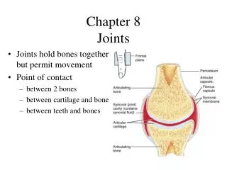
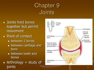
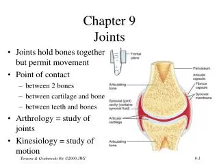
Joints (articulations) are unions or junctions between two or more bones or rigid parts of the skeleton. • Joints exhibit a variety of forms and functions. It is the fact that, whether or not movement occurs between them, it is still called a joint. • Some joints have no movement, others allow only slight movement, and some are freely movable.
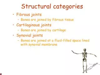
Classification of Joints Joints are classified according to the tissues that lie between the bones: Fibrous joints Cartilaginous joints Synovial joints
Fibrous joints • The bones are united by fibrous tissue. • The amount of movement occurring at a fibrous joint depends in most cases on the length of the fibers uniting the articulating bones. • The sutures of the cranium are examples of fibrous joints. • These bones are close together, either interlocking along a wavy line or overlapping.
A syndesmosis type of fibrous joint unites the bones with a sheet of fibrous tissue, either a ligament or a fibrous membrane. • Consequently, this type of joint is partially movable. • The interosseous membrane in the forearm is a sheet of fibrous tissue that joins the radius and ulna in a syndesmosis.
A dentoalveolarsyndesmosis (gomphosis or socket) is a fibrous joint in which a peglike process fits into a socket articulation between the root of the tooth and the alveolar process of the jaw. • Mobility of this joint (a loose tooth) indicates a pathological state affecting the supporting tissues of the tooth.
Cartilaginous joints The bones are united by hyaline cartilage or fibrocartilage. In primary cartilaginous joints, or synchondroses, the bones are united by hyaline cartilage, which permits slight bending during early life.
Secondary cartilaginous joints, or symphyses, are strong, slightly movable joints united by fibrocartilage. The fibrocartilaginous intervertebral discs between the vertebrae consist of binding connective tissue that joins the vertebrae together.
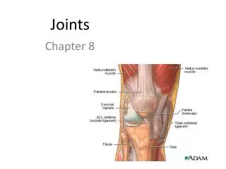
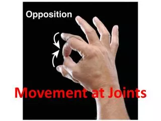
Synovial joints • The bones are united by a joint (articular) capsule (composed of an outer fibrous layer lined by a serous synovial membrane) spanning and enclosing an articular cavity. • Synovial joints are the most common type of joints and provide free movement between the bones they join. • They are joints of locomotion, typical of nearly all limb joints.
This type of joints has three common features: • Joint cavity: The joint cavity of a synovial joint, like the knee, is a potential space that contains a small amount of lubricating synovial fluid, secreted by the synovial membrane. • Articular cartilage: The articular surfaces are covered by hyaline cartilage
Articular capsule: This structure surrounds the joint and formed of two layers. Inside the capsule, articular cartilage covers the articulating surfaces of the bones; all other internal surfaces are covered by synovial membrane. • Fibrous capsule • Synovial membrane