Download
1 / 1
10 likes | 99 Vues
Xiaoming Zheng and Trachette Jackson Department of Mathematics, University of Michigan. A Multiscale Model of Cell Elongation, Proliferation and Quiescence Transition in Angiogenesis. Spring model of the tip cell elongation in a vessel:. Combined algorithm.

E N D
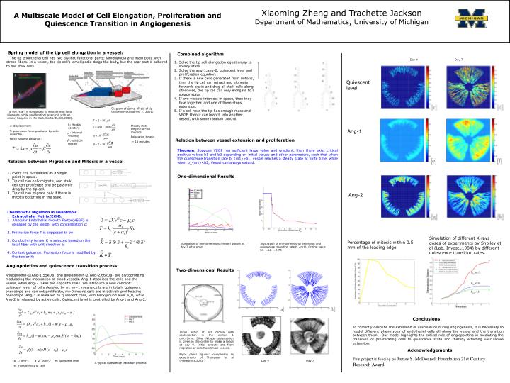
Xiaoming Zheng and Trachette Jackson Department of Mathematics, University of Michigan A Multiscale Model of Cell Elongation, Proliferation and Quiescence Transition in Angiogenesis Spring model of the tip cell elongation in a vessel: Combined algorithm The tip endothelial cell has two distinct functional parts: lamellipodia and main body with stress fibers. In a vessel, the tip cell’s lamellipodia drags the body, but the rear part is adhered to the stalk cells. Day 4 Day 7 Solve the tip cell elongation equation,up to steady state. Solve the ang-1,ang-2, quiescent level and proliferation equation. If there is new cells generated from mitosis, then the tip cell can retract and elongate forwards again and drag all stalk cells along, otherwise, the tip cell can only elongate to a steady state. If two vessels intersect in space, then they fuse together, and one of them stops extension. If a cell near the tip has enough mass and VEGF, then it can branch into another vessel, with some random control. Quiescent level Diagram of spring model of tip cell(Munevar,Biophys. J., 2001) Tip cell (star) is specialized to migrate with long filaments, while proliferation(green cell with an arrow) happens in the stalk(Gerhardt,JCB,2003). k: Hook’s constant : internal viscosity :cell-ECM friction u: displacement Steady state length=40~50 microns Relaxation time is ~ 10 minutes Ang-1 T: protrusion force produced by actin assembly. Force balance equation: Relation between vessel extension and proliferation Theorem. Suppose VEGF has sufficient large value and gradient, then there exist critical positive values b1 and b2 depending on initial values and other parameters, such that when the quiescence transition rate b_{m1}>b1, vessel reaches a steady state at finite time, while when b_{m1}<b2, Vessel can always extend. Relation between Migration and Mitosis in a vessel Every cell is modeled as a single point in space. Tip cell can only migrate, and stalk cell can proliferate and be passively drag by the tip cell. Tip cell can migrate only if there is mitosis occurring in the stalk. One-dimensional Results Ang-2 Chemotactic Migration in anisotropic Extracellular Matrix(ECM): Vascular Endothelial Growth Factor(VEGF) is released by the lesion, with concentration c: 2. Protrusion force T is supposed to be 3. Conductivity tensor K is selected based on the local fiber with unit direction a: 4. Contact guidance: Protrusion force is modified by the tensor K: Simulation of different X-rays doses of experiments by Sholley et al (Lab. Invest.,1984) by different quiescence transition rates. Percentage of mitosis within 0.5 mm of the leading edge Illustration of one-dimensional vessel growth at day 7 after onset. Illustration of one-dimensional extension and quiescence transition rate b_{m1}. Critical valus b1~=b2~=0.73 Angiopoietins and quiescence transition process Two-dimensional Results Angiopoietin-1(Ang-1,55kDa) and angiopoietin-2(Ang-2,66kDa) are glycoproteins modulating the maturation of blood vessels. Ang-1 stabilizes the cells and the vessel, while Ang-2 takes the opposite roles. We introduce a new concept: quiescent level of cells denoted by m: m=1 means cells are in totally quiescent phenotype and can not proliferate, m=0 means cells are in actively proliferating phenotype. Ang-1 is released by quiescent cells, with background level a_0, while Ang-2 is released by active cells. Quiescent level is controlled by Ang-1 and Ang-2. Conclusions To correctly describe the extension of vasculature during angiogenesis, it is necessary to model different phenotypes of endothelial cells all along the vessel and the transition between them. Our model highlights the critical role of angiopoietins in mediating the transition of proliferating cells to quiescence state and thereby effecting vasculature extension. Initial setup of rat cornea with cauterization in the center. 1 unit=2mm. Silver Nitrate cauterization is given in the center to make a lesion at day 0. Initial sprouts are from migration of cells from limbal vessels. Right panel figures: comparison to experiments of Thompson et al (Proteomics,2003 ) Acknowledgements This project is funding byJames S. McDonnell Foundation 21st Century Research Award. a_1: Ang-1 a_2: Ang-2 m: quiescent level e: mass density of cells Day 4 Day 7 A typical quiescence transition process