Download
1 / 39
920 likes | 2.06k Vues
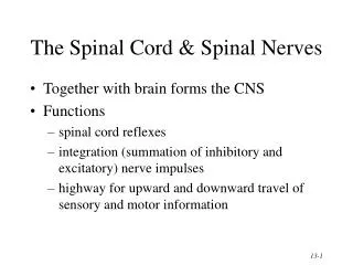
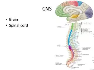
Chapter 13 The Spinal Cord & Spinal Nerves. Together with brain forms the CNS Functions spinal cord reflexes integration (summation of inhibitory and excitatory) nerve impulses highway for upward and downward travel of sensory and motor information. Spinal Cord Protection.

E N D
Chapter 13The Spinal Cord & Spinal Nerves • Together with brain forms the CNS • Functions • spinal cord reflexes • integration (summation of inhibitory and excitatory) nerve impulses • highway for upward and downward travel of sensory and motor information
Spinal Cord Protection By the vertebral column, meninges, cerebrospinal fluid, and vertebral ligaments.
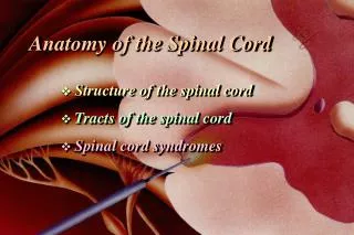
Structures Covering the Spinal Cord • Vertebrae • Epidural space filled with fat • Dura mater • dense irregular CT tube • Subdural space filled with interstitial fluid • Arachnoid = spider web of collagen fibers • Subarachnoid space = CSF • Pia mater • thin layer covers BV • denticulate ligs hold in place
External Anatomy of Spinal Cord • Flattened cylinder • 16-18 Inches long & 3/4 inch diameter • In adult ends at L2 • In newborn ends at L4 • Growth of cord stops at age 5 • Cervical enlargement • upper limbs • Lumbar enlargement • lower limbs
Inferior End of Spinal Cord • Conus medullaris • cone-shaped end of spinal cord • Filum terminale • thread-like extension of pia mater • stabilizes spinal cord in canal • Caudae equinae (horse’s tail) • dorsal & ventral roots of lowest spinal nerves • Spinal segment • area of cord from which each pair of spinal nerves arises
Spinal Cord & Spinal Nerves • Spinal nerves begin as roots • Dorsal or posterior root is incoming sensory fibers • dorsal root ganglion (swelling) = cell bodies of sensory nerves • Ventral or anterior root is outgoing motor fibers
Spinal tap or Lumbar Puncture • Technique • long needle into subarachnoid space • safe from L3 to L5 • Purpose • sampling CSF for diagnosis • injection of antibiotics, anesthetics or chemotherapy • measurement of CSF pressure
Gray Matter of the Spinal Cord Note: colors in reverse due to staining of tissue • Gray matter is shaped like the letter H or a butterfly • contains neuron cell bodies, unmyelinated axons & dendrites • paired dorsal and ventral gray horns • lateral horns only present in thoracic spinal cord • gray commissure crosses the midline • Central canal continuous with 4th ventricle of brain
White Matter of the Spinal Cord • White matter covers gray matter • Anterior median fissure deeper than Posterior median sulcus • Anterior, Lateral and Posterior White Columns contain axons that form ascending & descending tracts
Tracts of the Spinal Cord • Function of tracts • highway for sensory & motor information • sensory tracts ascend • motor tracts descend • Naming of tracts • indicates position & direction of signal • example = anterior spinothalamic tract • impulses travel from spinal cord towards brain (thalamus) • found in anterior part of spinal cord
Location of Tracts inside Cord • Motor tracts Sensory tracts • pyramidal tract (corticospinal) ---spinothalamic tract • extrapyramidal tract ---posterior column • ---spinocerebellar
Function of Spinal Tracts • Spinothalamic tract • pain, temperature, deep pressure & crude touch • Posterior columns • proprioception, discriminative touch, two-point discrimination, pressure and vibration • Direct pathways (corticospinal & corticobulbar) • precise, voluntary movements • Indirect pathways (rubrospinal, vestibulospinal) • programming automatic movements, posture & muscle tone, equilibrium & coordination of visual reflexes
Spinal Reflexes • Automatic response to change in environment • Integration center for spinal reflexes is gray matter of spinal cord • Examples • somatic reflexes result in skeletal muscle contraction • autonomic (visceral) reflexes involve smooth & cardiac muscle and glands. • heart rate, respiration, digestion, urination, etc • Note: cranial reflexes involve cranial nerves
Reflex Arc • Specific nerve impulse pathway • 5 components of reflex arc • receptor • sensory neuron • integrating center • motor neuron • effector • 4 important somatic spinal reflexes • stretch, tendon, flexor(withdrawal) & crossed extensor reflexes
Stretch Reflex (patellar reflex) • Monosynaptic,ipsilateral reflex arc • Prevents injury from over stretching because muscle contracts when it is stretched • Events of stretch reflex • muscle spindle signals stretch of muscle • motor neuron activated & muscle contracts • Brain sets muscle spindle sensitivity as it sets muscle tone (degree of muscle contraction at rest) • Reciprocal innervation (polysynaptic- interneuron) • antagonistic muscles relax as part of reflex
Tendon Reflex • Controls muscle tension by causing muscle relaxation that prevents tendon damage • Golgi tendon organs in tendon • activated by stretching of tendon • inhibitory neuron is stimulated (polysynaptic) • motor neuron is hyperpolarized and muscle relaxes • Both tendon & muscle are protected • Reciprocal innervation (polysynaptic) • causes contraction of ipsilateral muscle group
Flexor (withdrawal) Reflex • Step on tack (pain fibers send signal to spinal cord • Interneurons branch to different spinal cord segments • Motor fibers in several segments are activated • More than one muscle group activated to lift foot off of tack
Crossed Extensor Reflex • Lifting left foot requires extension of right leg to maintain one’s balance • Pain signals cross to opposite spinal cord • Contralateral extensor muscles are stimulated by interneurons to hold up the body weight • Reciprocal innervation - when extensors contract flexors relax, etc
Clinical Considerations • Checking a patient’s reflexes may help to detect disorders/injury • Plantar flexion reflex -- stroke the lateral margin of the sole • normal response is curling under the toes • abnormal response or response of children under 18 months is called Babinski sign (upward fanning of toes due to incomplete myelination in child)
Spinal Nerves • 31 Pairs of spinal nerves • Named & numbered by the cord level of their origin • 8 pairs of cervical nerves (C1 to C8) • 12 pairs of thoracic nerves (T1 to T12) • 5 pairs of lumbar nerves (L1 to L5) • 5 pairs of sacral nerves (S1 to S5) • 1 pair of coccygeal nerves • Mixed sensory & motor nerves
Connective Tissue Coverings • Endoneurium = wrapping of each nerve fibers • Perineurium = surrounds group of nerve fibers forming a fascicle • Epineurium = covering of entire nerve • dura mater blends into it at intervertebral foramen
Endoneurium Perineurium Epineurium
Branching of Spinal Nerve • Spinal nerves formed from dorsal & ventral roots • Spinal nerves branch into dorsal & ventral rami • dorsal rami supply skin & muscles of back • ventral rami form plexus supply anterior trunk & limbs • meningeal branches supply meninges, vertebrae & BV
A Nerve Plexus • Joining of ventral rami of spinal nerves to form nerve networks or plexuses • Found in neck, arm, low back & sacral regions • No plexus in thoracic region • intercostal nn. innervate intercostal spaces • T7 to T12 supply abdominal wall as well
Cervical Plexus • Ventral rami of spinal nerves (C1 to C5) • Supplies parts of head, neck & shoulders • Phrenic nerve (C3-C5) keeps diaphragm alive • Damage to cord above C3 causes respiratory arrest
Brachial Plexus • Ventral rami from C5 to T1 • Supplies shoulder & upper limb • Passes superior to 1st rib & under clavicle • Axillary n. = deltoid & teres m. • Musculocutaneous n. = elbow flexors • Radial n. = shoulder & elbow extensors • Median & ulnar nn. = flexors of wrist & hand
Clinical Correlations • Erb-Duchene palsy • waiter’s tip position • fall on shoulder • Radial nerve injury • improper deltoid injectionor tight cast • wrist drop • Median nerve injury • numb palm & fingers; inability to pronate & flex fingers • Ulnar nerve injury (clawhand) • inability to adduct/abduct fingers, atrophy of interosseus • Long thoracic nerve injury (winged scapula) • paralysis of serratus anterior, can’t abduct above horizontal
Lumbar Plexus • Ventral rami of L1 to L4 • Supplies abdominal wall, external genitals & anterior/medial thigh • Injury to femoral nerve causes inability to extend leg & loss of sensation in thigh • Injury to obturator nerve causes paralysis of thigh adductors
Branches of Lumbar Plexus • Notice: Femoral and Obturator nerves • Found anterior and medial to hip joint
Sacral Plexus • Ventral rami of L4-L5 & S1-S4 • Anterior to the sacrum • Supplies buttocks, perineum & part of lower limb • Sciatic nerve = L4 to S3 supplies post thigh & all below knee • Peroneal nerve injury produces foot drop or numbness • Tibial nerve injury produces calcaneovalgus (loss of function on anterior leg & dorsum of foot)
Branches of Sacral Plexus • Notice: Sciatic nerve origins
Sciatic Nerve Branches • Notice: Common Peroneal nerve and Tibial nerve behind the knee • Notice: Sciatica pain extends from the buttock down the leg to the foot • may be sign of herniated disc
Dermatomes & Myotomes • Each spinal nerve contains both sensory & motor nerve fibers • Dermatome • area of skin supplied by one spinal nerve • overlap prevents loss of sensation if one damaged • sensory anesthesia requires 3 spinal nerves to be blocked • Skin on face supplied by Cranial Nerve V
Dermatomes • Damaged regions of the spinal cord can be distinguished by patterns of numbness over a dermatome region • Infusing local anesthetics or cutting roots must be done over 3 adjacent spinal nerves. • Spinal cord transection • injury that severs the cord loss of sensation& motor control below the injury
Disorders • Neuritis • inflammation of nerves • caused by injury, vitamin deficiency or poison • Shingles • infection of peripheral nerve by chicken pox virus • causes pain, skin discoloration, line of skin blisters • Poliomyelitis • viral infection causing motor neuron death and possible death from cardiac failure or respiratory arrest