Download
1 / 27
300 likes | 668 Vues
Bone. 2007. Functions of Bone. Rigid skeleton supports the body Provides sites for attachment of muscles and organs Protective cover for soft organs Hemopoiesis (marrow cavities) Reservoir for body’s calcium supply

E N D
Bone 2007
Functions of Bone • Rigid skeleton supports the body • Provides sites for attachment of muscles and organs • Protective cover for soft organs • Hemopoiesis (marrow cavities) • Reservoir for body’s calcium supply • Calcium used for muscle contraction, blood clotting, cell membrane permeability, transmission of nerve impulses
Bone • Connective tissue with a mineralized matrix (calcium phosphate) • Matrix is dense and rigid, contains fibers of mainly Type I collagen. • Both the collagen and the ground substance become mineralized in bone.
Bone Matrix • Calcified matrix of fibers (Type I collagen) and ground substance (proteoglycans with chondroitin sulfate and keratan sulfate side chains, glycoproteins) • Organic component (osteoid)= type I collagen, highly cross-linked • Inorganic (mineral) component = crystals of calcium hydroxyapatite (calcium and phosphorus) • Hard – nutrients and metabolites cannot diffuse through the calcified matrix as done in cartilage. • Highly vascularized: • Canaliculi –small channels or canals between lacunae that allow osteocytes to exchange wastes and bring metabolites in; osteocytes send out cytoplasmic processes into the canaliculi and contact neighboring cells through gap junctions.
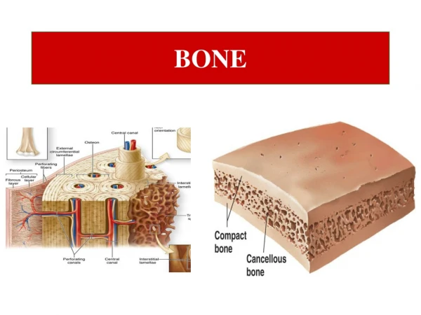
Structure of Bones • Periosteum – covers bone’s external surfaces and consists of an outer layer of dense, irregular collagenous (fibrous) CT and an inner layer of osteoprogenitor and osteoblast cells • Marrow – in central cavity of bone, hemopoietic tissue, red or yellow • Diaphysis – shaft • Epiphysis – articular ends
Epiphysis of an adult long bone Spongy (cancellous) bone Compact bone Compact bone
Spongy vs. Compact Bone • Spongy – composed of spicules or trabeculae of bone, forms network around bone marrow. Also known as cancellous bone. • Compact – dense bone, no cavities; found along the outer surface of shafts of long bone. Has lamellar structure of circumferential lamellae (on surfaces) and Haversian and interstitial lamellae. Haversian canal is space running through osteon with neurovascular bundle, osteoblasts and osteoprogenitor cells. Haversian canals of adjacent osteons connected by Volkmann’s canals.
Compact Bone – Low Powerhighly vascularized Haversian canal Volkmann’s canal
Bone Cells – 4 types • Osteogenic cells (osteoprogenitor cells) – undifferentiated stem cells, during development give rise to osteoblasts, line inner layer periosteum, haversian canals and endosteum. • Osteoblasts – immature bone cells; must secrete bone matrix to become mature. • Osteocytes – mature bone cells; trapped within lacunae of bone matrix they secrete around themselves. • Osteoclasts – large, multinucleate cells; found at the surfaces of bone where resorption and remodeling is taking place; motile cells; function to resorb bone in response to blood calcium levels and hormone stimuli. Howships lacunae – depressions made by osteoclasts as they etch away the bone.
Bones are dynamic structures! • Continually being resorbed and remodeled by osteoclasts • Hormones regulate amount of calcium in the blood • Parathyroid hormone – increases blood calcium levels; stimulates activity of osteoclasts and increases their numbers. • Calcitonin (from thyroid gland) – decreases blood calcium levels; reduces the numbers and activity of osteoclasts.
Bone Formation – 2 types • Intramembranous ossification – formation of bone directly from mesenchyme (embryonic connective tissue) with no cartilage precursor • Endochondral – formation of bone on cartilage scaffolding; bone eventually replaces cartilage.
Intramembranous Ossification • Occurs in flat bones of skull, mandible, maxilla. • Osteoblasts develop from connective tissue mesenchyme cells • Osteoblasts secrete bone matrix. • Form a network of bone spicules or trabeculae
Intramembranous Ossification From: Gartner and Hiatt, 2001
Intramembranous Ossification osteocytes osteoblasts From: Gartner and Hiatt, 2001
Intramembranous Ossification From: Gartner and Hiatt, 2001
Endochondral Ossification • Occurs in long and short bones, vertebrae • Bone is proceeded by temporary hyaline cartilage model that serves as a structural scaffold. • Bone tissue REPLACES the cartilage • Cartilage begins to calcify and causes chondrocytes to hypertrophy. • Cartilage calcifies so diffusion of nutrients to chondrocytes stops and they die. • Fragmented pieces of calcified cartilage serve as the framework to deposit the bone. • Osetoprogenitor cells and blood vessels from surrounding connective tissue penetrate and invade degenerating cartilage model. • The area/center of ossification expands, replacing cartilage with bone. • Cartilage of diaphysis replaced by bone except for at epiphyseal plate. • Primary and secondary centers of ossification.
Endochondral Ossification From: Gartner and Hiatt, 2001
Endochondral Ossification From: Gartner and Hiatt, 2001
Primary vs. Secondary Bone • Primary – immature or “woven” bon • First bone formed in fetal development or bone repair • Abundant osteocytes • Irregular bundles of collagen (Type I) • Secondary bone – mature or lamellar bone • Composed of concentric or parallel lamellae; osteocytes arranged in rows. • Canaliculi connect neighboring lacunae • Haversian canal system (osteon) • Cylinders of lamellae concentrically arranged around a central Haversian canal • Volkmann’s canals – connect one osteon to another; oriented perpendicular to Haversian canals.
Methods to Make Slides of Bone • Decalcified bone sections • Acid solution removes calcium salts and softens bone. • Bone can then be embedded and sectioned. • Osteocytes are distorted. • Ground bone sections • Saw bone in slices. • Grind piece down until thin enough to put on a slide. • Cells are destroyed and lacunae and canaliculi filled with bone debris.