Download
1 / 40
560 likes | 1.26k Vues
Autoimmune pancreatitis. Petr Dítě Dept. of Hepatogastroenterology Univ. Hospital Brno – Czech Republic. Incidence of Chronic Pancreatitis. Switzerland 1.2/100 000/year Poland 4.0/100 000/year Germany 7.4/100 000/year Czech Rep. 7.9/100 000/year

E N D
Autoimmunepancreatitis Petr Dítě Dept. of Hepatogastroenterology Univ. Hospital Brno – Czech Republic
Incidence of Chronic Pancreatitis Switzerland 1.2/100 000/year Poland 4.0/100 000/year Germany 7.4/100 000/year Czech Rep. 7.9/100 000/year Hungary 8.0/100 000/year Denmark 10.0/100 000/year Sweden 10.0/100 000/year Finland 23.0/100 000/year United States 5.7-7.6/100 000/year
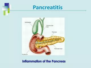
Chronic pancreatitis is a progressive inflammatory disease of the pancreas with irreversible damage of pancreatic tissueexocrine and endocrine insufficiency
TIGARO Classification T I GAR O -oxic-metabolic -diopathic -enetic -utoimmune -ecurrent acute pancreatitis -bstructive Etemed,Whitcomb, 2001
AUTOIMMUNE PANCREATITIS - chronic pancreatitis with distinct clinical, serological, histological and imaging features and it is involved in hyper- IgG4 group of diseases.
Autoimmunepancreatitis 1961 H. Sarles Chronic inflammatory sclerosis of the pancreas (Patients with jaundice, painful crises, hyperglobulinemia, no dilatation of pancreatic duct, lymphatic infiltration) • R. Waldram et al Chronic pancreatitis, sclerosing cholangitis and sicca sy in two siblings • S. Nakano et al Vanishing tumor of the abdomen in patient with Sjögren´s sy • K. Yoshida Concept of autoimmune pancreatitis 2001 B. Etemed, D. Whitcomb TIGARO classification
Epidemiology ofautoimmunepancreatitis Japan 21/451 4,6% Yoshida et al. Dig.Dis.Sci. 1995 Korea 17/315 5,4% Kim et al. Am.J.Gastroenterol. 2004 Italy 23/383 6,0% Parson et al. Pancreas 2003 Czech Rep. 9/185 4,8% Dite et al Best Practice and Res. Clin. Gastroent., 2008
Sex ang age onset of autoimmune pancreatitis Nishimori I. et al., Gastroent., 2007
Antibodies in patientswith AIP % Okazaki et al. J. Gastroent. 2001
CLINICAL DIAGNOSTIC CRITERIA FOR AIP 2006 • Diffuse or segmental narrowing of the MPD with irregular wall and diffuse or localized enlargement of the pancreas by imaging studies, such as abdominal US, CT, and magnetic resonance • High serum γ-globulin, IgG, or IgG4, or the presence of autoantibodies such as antinuclear antibodies and rheumatoid factor • Marked interlobular fibrosis and prominent infiltration of lymphocytes and plasma cells in the periductal area, occasionally with lymphoid follicles in the pancreas Diagnosis of AIP is established when criterion 1 and criterion 2 and/or 3 are fulfilled. However, it is necessary to exclude malignant diseases.
AUTOIMMUNE PANCREATITIS - SUBTYPES TYP 1 – LYMPHOPLASMATIC SCLEROSING PANCREATITIS – LPSP - PERIDUCTAL LYMPHOPLASMATIC INFILTRATE - HIGH AMMOUNT IgG4 - POSITIVE PLASMA CELLS - SWIRLING FIBROSIS - OBLITERATIVE VENULITIS TYP 2 – IDIOPATHIC DUCT-CENTRIC PANCREATITIS – IDCP (“non-alcoholic duct destructive pancreatitis“) - DUCTAL EPITHELIAL GRANULOCYTIC INFILTRATION DUCTAL DAMAGE OBLITERATION
COMPARISON OF TYPE 1 AND TYPE 2 AIP AIP,autoimmune pancreatitis, IgG4, immunoglobulin G4
CLINICAL PRESENTATIONS OF TYPE 1 AUTOIMMUNE PACREATITIS Clinical presentations of type I AIP Pancreatic Predominantly extra-pancreatic Acute Post-acute/late Biliary stricture, sclerosing cholangitis Obstructive jaundice Persistent mass Interstitial nephritis, renal failure Steatorrhea Pancreatitis Calcification, atrophy Retroperitoneal fibrosis with complications (e.g., ureteral obstruction) Steatorrhea Park, D.H. 2009
AUTOIMMUNE PANCREATITIS DIFFUSE FORM 77,0% (LIKE ACUTE PANCREATITIS) 23,0% FOCAL FORM (LIKE MALIGNANT LESION)
Scattergram of IgG4 values for patients with autoimmune pancreatitis and related diseases. PBC primary biliary cirrhosis, PSC primary sclerosing cholangitis Kawa et al., Gastroent., 2007
Usefulness of IgG4 in differentiating between pancreatic cancer and autoimmune pancreatitis Kawa et al., Gastroent., 2007
Abundant IgG4 – bearing plasma cell infiltration in patients with autoimmune pancreatitis and gastric ulcer Shinji, A. et al. Gastrointest. Endosc. 2004 23 ptswith AIP and 230 controlpatientsexamined by EGD In 8 ptswithautoimmunepancreatitisgastriculcerwasfound (34.8%). In controlgroupduring EGD wasgastriculcerfound in 31 pts (13.3%) = p.0007 Conclusion: AIP iscloselyassociatedwithgastriculcerwithabundant IgG4-bearing plasma cell infiltration
SET OF PATIENTS WITH AUTOIMMUNE PANCREATITIS(N = 10) Dítě,P. al 2010 One patient died during hospitalization – pancreatic cancer
Review of AIP cases with systemic extrapancreatic lesions Ohara et al, Pancreas 2005
AUTOIMMUNE PANCREATITIS IN PATIENTS WITH “IDIOPATHIC CHRONIC PANCREATITIS“ 66 PATIENTS WITH IDIOPATHIC CHRONIC PANCREATITIS /ICP/ AUTOIMMUNE DISEASE WAS PRESENT IN 10 PATIENTS (UC 5 pts, PSC 2 pst, Sjögren sy 1 pts, Hashimoto´s thyroiditis 1 pts, Graves disease 1 pts) POSITIVITY OF BIOCHEMICAL AND CLINICAL PARAMETRES – IN 40% CONCLUSION: CLINICAL OR BIOCHEMICAL AUTOIMMUNE STIGMATA ARE PRESENT IN 40% pts WITH ICP, AUTOIMMUNE MECHANISMS MAY BE FREQUENT IN ICP. Uzan,K.N. et al. Clin Gastroent. Hepatol. 2005
CHRONIC PANCREATITIS IN CHILDREN – AUTOIMMUNE ETIOLOGY? In the set of 31 children (age 3-18 years) • markers of AIP were found in 17 pts (41,5%) • Genetic markers 10 pts (32,5%) Oracz G. et al, Clin Gastroent Hepat 2006
Steroid therapy in patientswith AIP • Initial doses 30 – 40 mg per day for 2 – 4 weeks • The steroid therapy could be stopped after the period of 6 – 12 months. • Monitoring of laboratory and clinical symptoms are essential. • When AIP still appears after steroid therapy --- re-evaluation should be carried out taking pancreatic CARCINOMA into consideration! J.Jpn.Pacreas Soc., 2002
THERAPEUTIC OPTIONS IN PATIENTS WITH AIP • MAYO CLINIC – 11 WEEKS STEROIDS WITH TAPPERING DOSE 5 mg / WEEK • KIM – 1 mg/kg FOR 4 WEEKS AND TAPPERING THE DOSE 5 mg/WEEK • FRULLONI – 0,5 mg/kg FOR 4 WEEKS AND TAPPERING THE DOSE 5 mg/WEEK UNEFFECTIVE THERAPY – PANCREATIC CANCER
Long-term follow up study treating patients with AIP Steroid therapy 60 mg/day 1 pts 40 mg/day 1 pts Duration from 21 – 37 months 30 mg/day 7 pts 5 mg/day 1 pts Dose was tappered by 2.5 – 5.0 mg every two weeks Maintenance therapy: 5mg daily Follow up period – 4 years 6 monts Kamisawa et al. Pancreatology 2005
Long term therapypatientswith AIP - prognosis Kamisawa et al. Pancreatology 2005
AIP – ENDOCRINE AND EXOCRINE FUNCTION AFTER STEROID THERAPY 21 CASES AIP WITH STEROID THERAPY 10 CASES WITH EXOCRINE INSUFICIENCY • NORMALIZATION 8 • NO CHANGE 2 11 CASES WITH DIABETES MELLITUS • IMPROVEMENT 5 • AGGRAVATION 3 • NO CHANGE 3 Ito et al. 2007
Recurrenceofautoimmunepancreatitis Takayama et al (Amer.J.Gastroent. 2004) 42(11) 26% Wakabyashi et al.(Pancreas 2005) 36( 6) 17% Zamboni et al. Wirchow Arch. 2004) 22( 5) 23% Kim et al. (A.J. Gastroent. 2004) 17( 1) 6% Ramisawa et al. (J.Gastroenterol. 2007) 32( 2) 6%
THE THERAPY OF AIP RECCURENCE STEROID + AZATHIOPRINE 1mg/kg 2mg/kg FOR 11 WEEKS Mycophenolate or Rituximab are not effective S.CHari Abstr. DDW 2009
AUTOIMMUNE PANCREATITIS VS PANCREATIC CANCER - RADIOLOGIC IMAGING Kim et al., 2004
Usefulness of IgG4 in differentiating between pancreatic cancer and autoimmune pancreatitis Kawa et al., Gastroent., 2007
COMPARISON OF SUBJECTS WITH AIP AND PANCREATIC CANCER Ghazele A. et al. 2007
CONCLUSION AND PRACTICE POINTS • AIP IS NOT FREQUENT DISEASE IN EUROPE, MORE FREQUENT IN ASIA. • CLINICAL SYMPTOMS ARE USUALLY MILD (MOSTLY ABDOMINAL “DISCOMFORT“ WITHOUT PAIN ATTACKS) • IN CT, EUS OR US-DIFFUSE ENLARGEMENT OF PANCREAS (sausage pancreas), IN NMR-CP OR ERCP IRREGULAR NARROWING OF THE MAIN PANCREATIC DUCT ARE TYPICAL • PRESENCE NON-SPECIFIC ANTIBODIES IN BLOOD SERUM AND INCREASED LEVEL OF IgG AND IgG4 IN SERUM AND TISSUE • ASSOCIATION WITH OTHER AUTOIMMUNE DISEASE-TYP1 AIP • PANCREATIC CALCIFICATIONS AND/OR CYSTOIDS ARE NOT FREQUENT • THERAPY WITH STEROIDS IS EFFECTIVE • IN DIF. DG DIAGNOSIS AIP VS PANCREATIC CANCER – EUS GUIDED BIOPSY IS FUNDAMENTAL PROCEDURE