Download

1 / 52
520 likes | 663 Vues
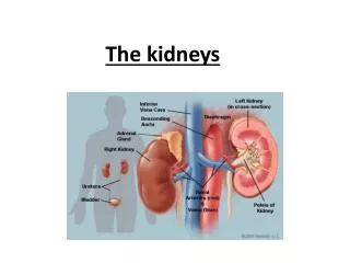
DEVELOPMENT OF the Kidneys and Ureters. Dr Rania Gabr. Objectives. 1- Describe the stages of development of the kidneys and ureters 2-Discuss the congenital anomalies of the kidneys and ureters. Stages of Development of the kidney:.

E N D
DEVELOPMENT OFthe Kidneys and Ureters Dr Rania Gabr
Objectives • 1- Describe the stages of development of the kidneys and ureters • 2-Discuss the congenital anomalies of the kidneys and ureters
Stages of Development of the kidney: • Human kidney is developed from the intermediate mesoderm and passes through 3 stages : 1- Pronephros 2- Mesonephros 3- Metanephros
PRONEPHROS • Appears at 4th week. In the cervical region, the cranial part of the intermediate mesoderm is segmented into 7 cell clusters called “nephrotomes”. • Those become cavitated to form 7 “pronephric tubules”. • Each tubule has 2 ends: 1- The lateral end:joins to form pronephric duct. 2-The medial end: joins the coelomic cavity.
The lowerend of the pronephricducts join the cloaca. • Fate of the Pronephros: 1-“Pronephric tubules: and proximal (cranial)part of its duct degenerate 2-The caudal part of the duct persists as the “Mesonephric duct”.
MESONEPHROS • It appears at the 6th week as 2 bulges on posterior abdominal wall forming ovoid mesonephric ridges. • It develops in the middle part of the (Intermediate mesoderm) that lies in thethoracic and upper lumbar region. • It is divided into segments which become canalised to form the “mesonephric tubules”.
The Medialends form the ( internal glomeruli ) while , • the lateral ends join the mesonephricductwhich opens in the primitive urogenital sinus.
Fate of the Mesonephros • 1- Tubules: - In Males: form the “vasa efferentia”. • Females:form epoophoron and paroophoron. 2- Duct: • Males: 1-Epididymis, 2-vas deferens, 3-seminal vesicle, 4-ejaculatory duct, 5-ureter trigone of urinary bladder and 6- the upper part of the posterior wall of the urethra.
In Females: 1-Duct of epoophoron (Gartener`s duct), 2-ureter and 3- trigone of urinary bladder. At the 8th week most of mesonephric tubules degenerate.
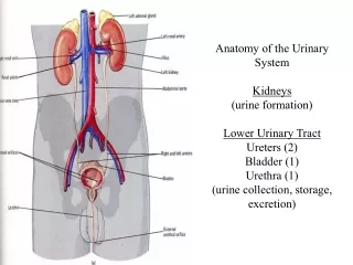
KIDNEY AND URETER • The human kidney develops from 2 main parts: 1- The ureteric bud, and 2- The Metanephros
The ureteric bud • The ureteric bud arises as an outgrowth of the mesonephric duct close to the entrance of the duct at the cloaca • The ureteric bud is responsible for the development of the collecting system
Ureter: • The ureteric bud elongates dorso-cranially to be in contact with the metanephroswhich will form the metanephric cap. • The upper end of the ureter divides to form 2 – 3 major calyces, which further divide into many minor calyces which divide into collecting tubules.
Each collecting tubule will be covered with a piece of the metanephric cap which form renal vesicles which form the rest of the nephron except the collecting tubules which is developed from dividing ureteric bud. • Collecting tubules communicate with the rest of the nephron.(canalization)
KIDNEY Metanephros: • It is the caudal part of the intermediate mesoderm in the pelvic cavity. • It forms the metanephric cap (blastema) which divides into small masses following divisions of the ureteric bud. • Each mass is called renal vesicles.
Each vesicle will form: 1- Bowman’s capsule, 2- proximal convoluted tubule, 3- loop of Henle and 4- distal convoluted tubule.
Changes of the external features of the developing kidney: • 1- Ascent of the kidney: It ascends from pelvic cavity to its adult site in the lumbar region on the posterior abdominal wall. This is done by the dorso-cranial elongation of the ureter pushing the kidney.
2- Change of the blood supply: During its ascent, the arterial supply of the kidney shifts to the nearest main artery . 1- First it is supplied by the Median sacral artery, then 2- Internal iliac, 3-Common Iliac, and finally 4- Abdominal aorta
3- Loss of fetal lobulation: • Thesurface of the kidney becomes smooth
4- Change of the direction of the hilum: from anterior to medial.
CONGENITAL ANOMALIESA- KIDNEY • 1- Renal agenesis:Unilateral or bilateral.
2- Congenital polycystic kidney:due to failure of communication between the collecting tubules and rest of the nephron.
4- Horse shoe kidney:due to fusion between lower ends of both kidneys. Ascent is arrested at level of L3 vertebra.
5- Rosette kidney: Due to fusion of the upper and lower poles
6- Persistence of fetal lobulation: The surface of the kidney shows lobulations.
7- Ectopic kidney: abnormal site of the kidney. • 8- Abnormal rotation Of the kidney: Rarely, the kidney shows lateral rotation instead of medial Rotation or faces anteriorly
9- Aberrant renal artery: An additional artery that supply the kidney. Might enter from the upper or lower poles. It is usually a non degenerated Artery during ascent.
CONGENITAL ANOMALIESB- Ureter • 1- Bifid ureter: Splitting of the upper part of the ureter.
2- Double ureter and Ectopic ureter:Two separate ureters due to formation of 2 ureteric buds. • One will open normally in the bladder, The other (Ectopic ureter): Male: Opens in the urethra Female: Opens either in : a- the vestibule of the vagina, or b- in the urethra