Download
1 / 65
680 likes | 863 Vues
Approach to Paroxysmal Supraventricular Tachycardias. M.V.Jorat MD 1389. Definition. Narrow QRS complex supraventricular tachycardia (SVT) is a tachyarrhythmia with a rate more than 100 beats/min and a QRS duration of less than 120 milliseconds.

E N D
Approach to Paroxysmal Supraventricular Tachycardias M.V.Jorat MD 1389
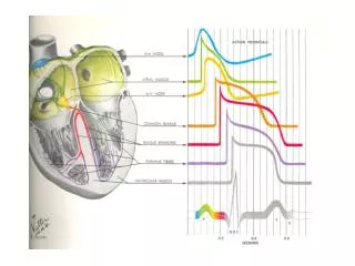
Definition • Narrow QRS complex supraventricular tachycardia (SVT) is a tachyarrhythmia with a rate more than 100 beats/min and a QRS duration of less than 120 milliseconds. • A narrow QRS complex (<120 msec) reflects rapid activation of the ventricles via the normal His-Purkinje system, which in turn suggests that the arrhythmia originates above or within the atrioventricular (AV) node (ie, a supraventricular tachycardia).
Differential Diagnosis of Narrow QRS Complex Tachycardias • Sinus tachycardia • Inappropriate sinus tachycardia • Sinoatrial nodal reentrant tachycardia (SNRT) • Atrial tachycardia (AT) • Multifocal AT • Atrial fibrillation (AF) • Atrial flutter (AFL) • Junctional ectopic tachycardia (JET) • Nonparoxysmaljunctional tachycardia • Atrioventricular nodal reentrant tachycardia (AVNRT) • Atrioventricular reentrant tachycardia (AVRT)
Tachycardia Narrow QRS tachycardia Wide QRS Tachycardia Regular Irregular AV node independent AV node dependent AF, AT/AFL with variable AV conduction, multifocal AT Sinus tachycardia Junctional tachycardia AT AFL AVNRT AVRT
Paroxysmal SVT is the term generally applied to intermittent SVT other than AF, AFL, and multifocal AT. • The major causes are: • AVNRT (approximately 50% to 60% of cases), • AVRT (approximately 30% of cases), • and AT (approximately 10% of cases). • The estimated prevalence in the normal population: 2.25/1,000 • Incidence: 35/100,000 person-years. • Age: In the absence of structural heart disease it can present at any age but most commonly first presents between ages 12 and 30 years. • Sex: Females/ males: 2
Proportion of paroxysmal supraventricular tachycardia mechanism by age This trend is similar for both gender
Initial Evaluation • History, physical examination, and an ECG constitute an appropriate initial evaluation of paroxysmal SVT. • Clinical symptoms are not usually helpful in distinguishing different forms of paroxysmal SVT. • Different methods of electrocardiographic evaluation helps for detection of arrhythmias.
Clinical Presentation • Onset and offset: • Abrupt or gradual (warm up and cool down) onset and termination of palpitation • Abnormal automacity or reentrant mechanism • Duration: from minutes to several hours • Commonly is associated with dizziness • Rapid ventricular rates can be associated with: • Dyspnea, weakness, angina, or even frank syncope, and can at times be disabling. • Neck pounding (AVNRT) • Patients often learn to use certain maneuvers such as carotid sinus massage or the Valsalva maneuver to terminate the arrhythmia
Physical examination • In patients without structural heart disease, the physical examination is usually remarkable only for a rapid, regular heart rate. • At times, because of the simultaneous contraction of atria and ventricles, cannon A waves can be seen in the jugular venous waveform. • In patients with an AT exhibiting AV block, usually of the Wenckebach type, the ventricular rate is irregular.
An echocardiographic examination should be considered in patients with documented sustained SVT to exclude the possibility of structural heart disease. • Exercise testing is less often useful for diagnosis unless the arrhythmia is clearly triggered by exertion. • Invasive EP testing with subsequent catheter ablation may be used for diagnosis and therapy in cases with a clear history of paroxysmal regular palpitations.
Clinical history of palpitation 12 lead ECG in normal sinus rhythm Preexitation YES NO Suspected to AVRT Asses arrhythmia pattern by clinical history Irregular palpitation Suspected AF, MAT, AFL or AT by variable block Syncope Sustained regular palpitation Event recorder and follow up Refer to Electrophysiologist
12 lead ECG: 12-lead ECG only during tachycardia can be helpful for defining the mechanism of paroxysmal SVT. • Ambulatory 24-hour Holter recording may be used for: • Documentation of the arrhythmia in patients with frequent (i.e., several episodes per week) but self-terminating tachycardias. • A cardiac event monitor is often more useful than a 24-hour recording in patients with less frequent arrhythmias. • Implantable loop recorders can be helpful in selected cases with rare episodes associated with severe symptoms of hemodynamic instability (e.g., syncope).
Heart Monitoring Options 10 Seconds 12-Lead 2 Days Holter Monitor Typical Event Recorder 7 Days MCOT External Loop Recorder 30+ Days 36 Months ILR ILR = insertable loop recorder MCOT= mobile cardiac outpatient telemetry
External Mobile Cardiac Outpatient Telemetry (MCOT) ECG Recorder PDA stores ECG data and symptom status. Wireless transmission capability provided. Patient Indicates symptoms on PDA. Abnormal ECG transmitted automatically Monitor center receives, reviews and transmits data to physician. Pre-determined ‘urgency’ criteria determine timing of physician alerts Physician receives and acts upon data as medically appropriate Cardionet Inc., San Diego, CA
Insertable Loop Recorder (ILR) An ECG monitoring system that is implanted subcutaneously Capable of recording, storing, and if necessary remotely transmitting ECG signals Patient-activated and/or automatically-activated Longevity of current ILRs up to 36 months Indicated for Patients with unexplained syncope / TLOC Patients who experience transient symptoms that may suggest a cardiac arrhythmia Patients at increased risk of cardiac arrhythmias
Insertable Loop Recorders (ILR) • Reveal® system, • Medtronic Inc., Minneapolis, MN • manual/auto trigger • remote download (CareLink®) • Sleuth®, • Transoma Inc., St Paul, MN • manual/auto trigger • wireless data transmission • Confirm®, • St Jude Medical • St Paul, MN • manual/auto trigger • remote download
ILR Symptom-Rhythm Correlation:Case Examples Case: 56 year old woman with refractory syncope accompanied with ‘seizures’. Case: 65 year old man with syncope accompanied by brief retrograde amnesia. *Medtronic data on file
PR dispersion for determination of Preexitation Tracings from the 12-lead ECG illustrating the principle of PR dispersion. Lead II has the least pre-excited QRS complex with the longest PR interval (180 ms), whereas lead V5 is the most pre-excited with the PR interval of 100 ms. Thus, the PR dispersion was calculated as 180- 100=80 ms.
Use aVR for excludion of preexitation • Four patterns of the QRS complexes in lead V6 and corresponding patterns in lead aVR in the same patient. • Positive delta wave obscuring septal activation. • Isoelectricdelta wave resulting in pseudo-septal R wave. • Overt pre-excitation without any isoelectric line between P wave and positive delta wave. • Manifest septal Q wave.
Stepwise algorithm to exclude or confirm pre-excitation on a 12-lead ECG Europace (2010) 12, 119–123
Most SVTs are associated with a regular ventricular rate. • If the rhythm is irregular, the ECG should be scrutinized for discrete atrial activity and for any evidence of a pattern to the irregularity (e.g., grouped beating typical of Wenckebach periodicity). • If the rhythm is irregularly irregular (i.e., no pattern can be detected), the mechanism of the arrhythmia is either multifocal AT or AF.
Multifocal AT is an irregularly irregular atrial rhythm characterized by: • More than three different P wave morphologies, • The P waves separated by isoelectric intervals • Varying P-P, R-R, and PR intervals
Atrial Fibrillation • AF is characterized by rapid and irregular atrial fibrillatory activity and, in the presence of normal AVN conduction, by an irregularly irregular ventricular response. • P waves cannot be detected in AF, although coarse fibrillatory waves and prominent U waves can sometimes give the appearance of P waves. • The fibrillatory activity sometimes is so fine as to be undetectable.
Naroow QRS tachycardia Regular tachycardia? Visible P wave? AF, AT/AFL with variable AV conduction, multifocal AT Atrial rate>ventricular rate? AFL or AT Analyze RP interval RP<PR (short RP tachycardia) RP>PR (long RP tachycardia) RP interval <70 ms RP interval>70 ms AT, PJRT, atypical AVNRT AVRT AVNRT AT Typical AVNRT
Identification of the Atrial Activity • If the patient's rhythm is regular or has a clearly discernible pattern, the ECG should next be assessed for P waves (atrial activity). • The P waves may be easily discernible; however, frequently, comparison with a normal baseline ECG is needed and can reveal a slight alteration in the QRS, ST segment, or T or U waves, suggesting the presence of the P wave. • If the P waves cannot be clearly identified, carotid sinus massage or the administration of intravenous adenosine may help clarify the diagnosis. • These maneuvers may also terminate the SVT.
Carotid Sinus Massage • Temporary decrease in the atrial rate in patients with: • Sinus tachycardia • Automatic AT. • Slowing of AVN conduction and AVN block, which can unmask atrial electrical activity that is, reveal P waves or flutter waves in patients with: • AT • AFL
Carotid Sinus Massage 3. With some SVTs that require AVN conduction, especially AVNRT and AVRT, the transient slowing of AVN conduction can terminate the arrhythmia by interrupting the reentry circuit; • Less commonly, carotid sinus massage can cause some ATs to slow and terminate. 4. In some cases, no effect is observed.
Adenosine Administration • Adenosine results in slowing of the sinus rate and AVN conduction. • In the setting of SVT, the effects of adenosine are similar to those seen with carotid sinus massage.
For intravenous adenosine administration, the patient should be supine and should have ECG and blood pressure monitoring. • The drug is administered by rapid intravenous injection over 1 to 2 seconds at a peripheral site, followed by a normal saline flush. • The usual initial dose is 6 mg, with a maximal single dose of 18 mg. • If a central intravenous access site is used, the initial dose should not exceed 3 mg and may be as little as 1 mg. • Adenosine can precipitate AF and AFL because it shortens atrial refractoriness. • In patients with Wolff-Parkinson-White (WPW) syndrome and AF, adenosine can result in a rapid ventricular response that can degenerate into VF.
Use of adenosine for diagnosis and termination of regular SVTs, including AVRT, is appropriate as long as close patient observation and preparedness to treat potential complications are maintained.
Termination of the Arrhythmia. • Carotid sinus massage or adenosine can terminate the SVT, especially if the rhythm is AVNRT or AVRT. • A continuous ECG tracing should be recorded during these maneuvers, because the response can aid in the diagnosis. • Termination of the tachycardia with a P wave after the last QRS complex is most common in AVRT and typical AVNRT and is rarely seen with AT. • Termination of the tachycardia with a QRS complex is more common with AT, atypical AVNRT, and permanent junctional reciprocating tachycardia (PJRT). • If the tachycardia continues despite development of AV block, the rhythm is almost certainly AT or AFL; AVRT is excluded and AVNRT is very unlikely.
Atrial Rate • An atrial rate more than 250 beats/min is almost always caused by AFL. • However, overlap exists, and AT and AVRT can occasionally be faster than 250 beats/min. • AVRT tends to be faster than AVNRT and AT.
P Wave Morphology • P wave morphology identical to sinus P wave: • Sinus tachycardia • Inappropriate sinus tachycardia • Sinoatrial nodal reentrant tachycardia • AT arising close to the region of the sinus node. • An abnormal P wave morphology: • AVNRT (P wave is concentric) • AVRT (P wave can be eccentric or concentric) • AT (P wave can be eccentric or concentric) • AFL (lack of distinct isoelectric baselines between atrial deflections is suggestive of AFL but can also be seen occasionally in AT.
The P waves may not be discernible on ECG, which suggests: • Typical AVNRT • Less commonly, AVRT (especially in the presence of bundle branch block [BBB] contralateral to the BT).