Download
1 / 40
420 likes | 800 Vues
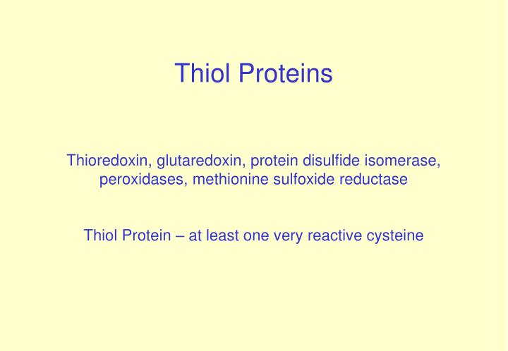
Thiol Proteins Thioredoxin, glutaredoxin, protein disulfide isomerase, peroxidases, methionine sulfoxide reductase Thiol Protein – at least one very reactive cysteine. Thiols in Biology. COO - COO -

E N D
Thiol Proteins Thioredoxin, glutaredoxin, protein disulfide isomerase, peroxidases, methionine sulfoxide reductase Thiol Protein – at least one very reactive cysteine
Thiols in Biology COO - COO - H-C-CH2-SH H-C-CH2-S- + H+ pKa = 8,37 NH3+ NH3+ cysteine pH = 8,37: [RSH] = [RS-] thiol thiolate pH < 8,37 [RSH] > [RS-] pH > 8,37 [RSH] < [RS-]
N H O H O 3 O O C C H C H C H C N H C C N H C H C 2 2 2 O O C H 2 S H c y s t e i n e g l y c i n e - g l u t a m a t e l Glutathione (GSH) pKa = 9,2 Low toxicity High intracellular concentrations (1-10mM)
Thiol/disulfide exchange reactions R1SH + R2SS R2 R1SS R2 + R2SH • central theme in biology • structure of proteins • Regulation of protein activity • (enzyme, transcription factors...) • -cellular redox homeostasis etc...
Rlg Rn Rn-S- + S-S S-S + Rlg-S- R R k nucleophilic agent leaving group Mechanism of thiol/disulfide exchange reactions nucleophilic substitution V = k[Rn-S- ] [ RSSRlg]
Thioredoxin (Trx) Dissulfide reductase - 12-13kDa - plants, animals, yeast , bacteria target trx trx H H target target trx trx Cys35-SH Cys32-S - Cys35-SH Cys32-S - S S S S Cys35-S Cys32-S - -S S -S S Cys35-S Cys32-S Cys35-S Cys32-S HS HS HS HS thioredoxin reductase NADPH + trx (-SS-) NADP + trx (-2SH) glutathione reductase NADPH + GSSG NADP + 2 GSH
Thioredoxin fold thioredoxin
Ribonucleotide Reductase, other target oxidized Grx GSSG (2-RSH) (RSSR) GSSG reductase NADPH,H+ Ribonucleotide Reductase, other target reduced Grx 2 GSH ) Glutaredoxin (Grx) 12-13kDa CXXC motifi (CPYC)
Trx and Grx targets yeast PAPS Tioredoxin reductase peroxidase Ribonucleotide Reductase Methionine ? ? sulphoxide ? reductase Oxidized Grx1 GSSG Trx1 Target Grx2 Trx2 (2 - RSH) (2 - RSH) Glr1, NADPH, H Trr1, NADPH, H + + Reduced Grx1 Trx1 Target Grx2 GSH Trx2 ( - SS - ) ( - SS - )
Methionine sulfoxide reductase Reviewed by Weissbach et al. (2002) Arch. Biochem. Biophys., 397:172.
Deglutathionylation by Grx monothiol mechanism Grx-S- + protein –SS G Grx – SS G + protein-SH Grx – SS G + GSH GSSG + Grx-S-
Protein Motif in active site Redox Potential (mV) TrxCys-Gly-Pro-Cys -270 GrxCys-Pro-Tyr-Cys -200 to –235 TryparedoxinCys-Por-Pro-Cys -249 Protein disulfide isomerase Cys-Gly-His-Cys -127 (PDI) DsbA Cys-Pro-His-Cys -125 Thiol/disulfide oxido-reductases
PDI and DsbA generate disulfide bonds in proteins PDI is in the endoplasmatic reticulum (ER) DsbA in periplasm (bacteria) GSH:GSSG in ER 1:1 to 3:1 (100:1 to 30:1 in cytoplasm)
Protein Oxidation States TrxCys-Gly-Pro-Cys-2 (thiol), -1 (disulfide) GrxCys-Pro-Tyr-Cys-2 (thiol), -1 (disulfide) TryparedoxinCys-Por-Pro-Cys-2 (thiol), -1 (disulfide) Protein disulfide isomerase Cys-Gly-His-Cys-2 (thiol), -1 (disulfide) (PDI) DsbA Cys-Pro-His-Cys -2 (thiol), -1 (disulfide) Glutathione Peroxidase Asn-Val-Ala-Ser-Lys-Cys -Gli -2 (thiol), 0 (sulfenic acid) non-selenium (GPx) Thiol proteins oxidation states
Prx = peroxiredoxin 2 RSH + H2O2 RSSR + 2 H2O 2 RSH + ROOH RSSR + ROH + H2O 197 residues - 25kDa Active site: cysteine 47 cysteine 170 – catalysis Netto et al.(1996) J. Biol. Chem., 271, 15315-15321.
Trivelli, X., Krimm, I., Ebel, C., Verdoucq, L., Prouzet-Mauléon, V., Chartier, Y., Tsan, P., Lauquin, G., Meyer, Y., Lancelin, J. (2003) Characterization of the yeast peroxiredoxin Ahp1 in its reduced and overoxidized inactive forms using NMR. Biochemistry42: 14139-49. Type A YPxDF[T/S]FVCPP[T/S]E[I/L/V] .....C-terminal VCrP Type B HPxDFTPVCPTTE Type C YPx[A/D]xTP[G/V] CPTx[Q/E]xCrx[F/L] Type D xP[G/A]A[F/Y][T/S][P/G]xCP[S/T]xxHxP Type E xP[D/S]DTxVCPxx[Q/S]x[K/R]
Trx = Prx = Gpx Amino acid sequence Prx and Trx have the same fold Trx Prx Choi et al. (1998) Nature Struct. Biol.5: 400 Gpx Prx
Peroxynitrite reductase activity of bacterial Prx Bryk, R., Griffin, P. & Nathan, C. Nature (2000), 407:211-215 Prx OONO - + H+ + Trx NO2 + H2O + Trx 106 M-1 s-1 SH S- S -- S
1-cys 2-cys típica 2-cys atípica
+ DTT (-SS-) 61 125 SH SH 61 125 61 125 61 125 SOH SH SS-DTT-SH SH SSG SH 61 125 S S + DTT ROOH + H2O fast Sulfenic acid Reduced protein disulfide GSSG 61 125 61 125 SO3H SH SO2H SH ROOH GSH GSH ROOH Glutathionylated protein Sulfonic acid (oxidation state = +4) Sulfinic acid (oxidation state = +2)
Versatility of Prx Prx (AhpC) as GSSG reductase Mutation = one amino acid insertion (Phe) Ritz et al (2001) Science294: 158
Crystal structure of oxydized decamer High concentrations Disulfides in yellow Thioredoxin fold: 2x (beta-alfa-beta)
2-Cys –Prx are also chaperons!! WT Delta prx1/prx2 Delta prx1/prx2 + pRS416/cTPxI Delta prx1/prx2 + pRS416/C47S/C170S Heat shock (43 C por 30’) plating
Proteins analyzed by SEC cTPxI,cTPxII 40-1000 kDa!! MW = 21,5 kDa (2-50 proteínas) Western blot 10% PAGE
Electron Microscopy (EM) FI fraction Particle diameter: 22-28nm
Electron Microscopy (EM) Two views: 409 “End on” (five fold symmetry) 170 “Double dot” FII fraction Particle diameter: 14 nm
R-SH P-S47 - S170-P R-SH P-S47H R-S47- P-S47OH INATIVA REATIVAINSTÁVEL peróxido P-S47O2H oligomerização
R-SH P-S47 - S170-P R-SH P-S47H R-S47- P-S47OH INATIVA REATIVAINSTÁVEL srx peróxido P-S47O2H oligomerização
Sulfiredoxin (Srx) – 13kDa Cys 84 – reactive site Homolog in humans Biteau et al. (2003), Nature 425: 980
Number of genes >>>>> Number of folds Trx Grx GSH transferase GSH peroxidase Prx Calsequestrins PDI Methionine sulfoxide reductase Trx fold
Trx X Cytochrome c Maturation Protein • (CMP) • - high amino acid sequence divergence • trx fold • Conserved residues involved in catalysis
Motif analysis Trx, CMPs and Prx common ancestor !!
Ohr = Organic Hydroperoxide Resistance protein • Deletion of Ohr gene: • Bacteria sensitive to organic peroxides (not H2O2!!) • Only organic peroxides induce transcription of Ohr gene New peroxiredoxin from Xylella fastidiosa ?
ROS RNS pathogen PLANT
A. B. TBHP (200 M) H2O2 (200 M) Ohr (2 ng/ul) Ohr (50 ng/ul) Ohr is a thiol — dependent peroxidase Cussiol J.R.R., Alves S.V., Oliveira M.A. e Netto L.E.S. (2003) J. Biol. Chem , 278, 11570—11578
Ohr - Xylella fastidiosa Tratada com t-BOOH (1mM/RT/1h) Tampão – Tris-Cl pH=8,5 0,1MPrecipitante – PEG 4000 25% Complete dataset at 1.9 Å Oliveira et al., (2004) Acta Crystall. D60, 337
Ohr structure No trx fold – alfa-beta fold Ohr Prx GSH px. New class of thiol dependent peroxidase