Download
1 / 21
210 likes | 633 Vues
54- year - old woman with newly diagnosed esophageal cancer. Associate Professor , Dr. Umut Kefeli, Kocaeli University School of Medicine Department of Medical Oncology. 8 th International Gastrointestinal Cancer Conference 07.12.2018. Case Presentation. 54-year-old ♀ 2 children

E N D
54-year-oldwomanwithnewlydiagnosedesophagealcancer AssociateProfessor, Dr. Umut Kefeli, Kocaeli University School of MedicineDepartment of MedicalOncology 8th International GastrointestinalCancer Conference 07.12.2018
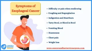
CasePresentation 54-year-old ♀ 2 children ECOG PS 0 Hypothyroidism No history of smoking,alcohol Dysphagia
Upper GIS endoscopy: Thoracalesophagealstricture at nearly 35 cm fromtheincisors. • Thorax CT: 40 mm longirregularwallthickening at distalthoracicesophagus, nearlytotallyobliteratingthelumenwhichsuggests an esophagealmass .
ThoraxCT: - Mass lesioncausesindentations of theright pulmonaryarteryandtheposteriorcontour of theleftatrium. - No clearfatplanebetweendescending aorta andthemasslesion. - Prevascular, paratrachealandrightparaesophageal, 14x7 mm. lymphadenopathies. - Right lungparenchymal, upto4.5 mm three pulmonarynodulesand2 mm subpleural noduleat leftlowerlobe.
PET/CT: - 37 mm long, infracarinalesophagealwallthickening (SUVmax:32) - Right upperparatracheal, bilaterallowerparatracheal, leftprevascular, precarinal, subcarinalandbilateralhilarminimallyincreased hypermetabolic lymphadenopathies (SUVmax:4.9). - No FDG involvement in thepulmonaryparenchymaandabdominopelviclymphnodes.
Histological examination of the endoscopic biopsy specimen demonstrated moderately-differentiated squamouscell carcinoma . • T4(?), N(+), M(-), clinicalstage IVAesophageal cancer was diagnosed.
WHAT WOULD BE YOUR SUGGESTION? A) Neoadjuvant chemotherapy B) Definitive chemoradiation C) Preoperative chemoradiation D) Surgery
Treatedwithdefinitivechemoradiation : 5-Fluoruracil andCisplatinand RT to50.4 Gy. • Patient is re-evaluatedwith PET/CT. • PET/CT reported a nearly total improvement in esophagealwallthickeningwith a total metabolicresponseandminimallyincreased FDG uptake in allmediastinalstationssuggestingreactivedisease. • Thorax CT reported a wallthickening of 8 mm in itsthickestplacewithreversal of esophagealdilatationandmultiplemediastinallymphnodesupto 10 mm diameter.
WHAT WOULD BE YOUR SUGGESTION? A) Surveillance B) Surgery C) Palliativemanagement D) Chemotherapy
Total esophagectomy + leftthoracotomy + witzelljejunostomy (16.11.2017): I. Total esophagectomy + subtotalgastrectomymaterial - Connectivetissuedevelopment, frequentcoagulationnecrosisareas - 1 lymphnode: carcinomametastasis - 9 lymphnodes: reactivehyperplasia II. Subcarinallymphnode: anthracosis, granulomas III. İnferiorligamentbiopsy: histiocytosis, anthracosis, granulomas No tumorsseen at surgicalborders .
Capecitabineand oxaliplatin - Capecitabine1000 mg/m2 PO BID on Days 1–14 - Oxaliplatin130 mg/m2 IV on Day 1 - Cycled every 21 days • After 6 cycles of adjuvantchemotherapy, patient is re-evaluatedwith a PET/CT whichshowedno FDG uptake( 04.05.2018 ) • There wasnopathologicallesion in ThoraxCTs (04.11.2018).
CasePresentation -2 79-year-old ♂ 2 children ECOG PS 2 GERD , COPD, CAD Persistentdysphagia
Esophagography (2007): marked dilatation of the esophagus, to about 2 cm in diameter, proximal to the gastroesophageal junction, which was diagnosed as achalasia of the esophagus. • Endoscopic balloon dilatation was performed threetimes from May 2007to 2015.
In September 2015, routine follow-up upper GI endoscopy revealed a shallow depressed lesion(0-IIc) in the proximal esophagus located 25 cm fromtheincisors. • The biopsy findings indicated moderately differentiated squamous cell carcinoma. • Endoscopic ultrasound (EUS) was performed andrevealed a blurring and thickening of the third layer (submucosal layer) but not fourthlayer. • There werenolymphnodesseen. • No metastases on a computed tomography (CT) scan or a positron emission tomography (PET)-CT scan.
ESD was made difficult by bleeding from abundant microvessels in the submucosal layer. • The lesion was excised and pathological findings revealed partial thickening of the mucosa and squamous cell carcinoma (0-IIc, 41 x 57 mm, depth T1a-EP(M1), ly0, v0, pHM0, pVN0).
In May 2017, routine follow-up upper GI endoscopy againshowed a shallow depressed lesion (0-IIc) in the upper esophagus. • An ESD is performedagain. • The pathological examination revealed squamous cell carcinoma (0-IIc, 21 x 13 mm, depth T1b(SM2), ly0, v2, pHM0, pVM0). • Patient refusedsurgery.
WHAT WOULD BE YOUR SUGGESTION? A) Surveillance B) Surgery C) Definitivechemoradiation
Treatedwithdefinitivechemoradiation : Capecitabineandoxaliplatin and RT to 50.4 Gy. - Oxaliplatin 85 mg/m2 IV on Days 1, 15, and 29for 3 doses - Capecitabine 625 mg/m2 PO BIDon Days 1–5 weekly for 5 weeks. • Therewere no pathologicallesions in follow-upimagings. • Since then, the patient has had no recurrence.