Download
1 / 24
240 likes | 315 Vues
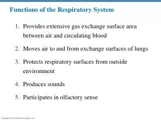
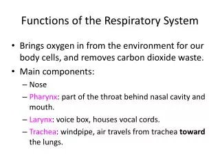
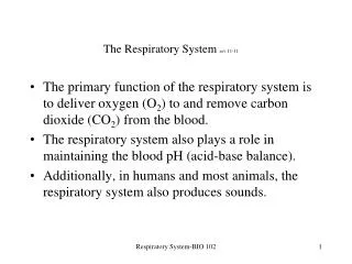
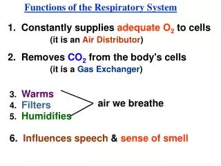
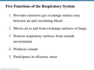
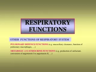
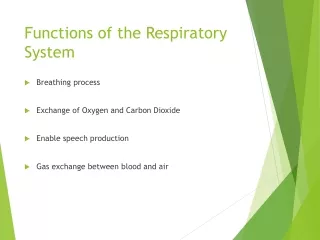
Respiratory System rev 12-09 Performs 2 basic functions: Air distribution Gas exchange—oxygen and carbon dioxide Process of respiration is another important homeostatic mechanism; it enables our cells to function effectively Respiratory system filters, warms and humidifies the air we breathe.

E N D
Respiratory Systemrev 12-09 • Performs 2 basic functions: • Air distribution • Gas exchange—oxygen and carbon dioxide • Process of respiration is another important homeostatic mechanism; it enables our cells to function effectively • Respiratory system filters, warms and humidifies the air we breathe Respiratory System BIO 006
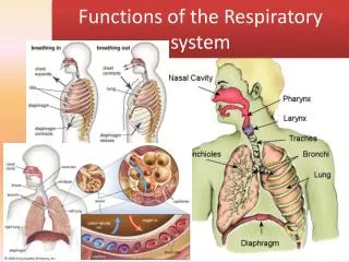
Upper respiratory tract— • nose, pharynx, and larynx • Lower respiratory tract— • trachea, bronchial tree, and lungs including the alveoli Respiratory System BIO 006
Respiratory Mucosa • Specialized membrane that lines the air distribution tubes in the respiratory tree • Covered with mucus and ciliated cells • More than 125 ml of mucus produced each day forms a “mucous blanket” over much of the respiratory system • Helps to clean, warm and humidify air we breathe • Mucus moves only toward the pharynx except in smokers • Cigarette smoke paralyzes the cilia and this causes smoker’s cough Respiratory System BIO 006
Nose or External Nares • Structure • Nasal septum separates interior of nose into two nasal cavities which are lined by mucous membrane • Nerve endings responsible for the sense of smell are located in the nasal mucosa • Functions: Warms and moistens inhaled air • Frontal, maxillary, sphenoidal, and ethmoidal sinuses drain into nose • Nasal mucosa also lines the sinuses and sinusitis can often develop from colds • 2 ducts from the lacrimal sacs drain into the nasal cavity Respiratory System BIO 006
Pharynx • Structure • Also called the throat • Both nasal cavities, the mouth, the esophagus, the larynx, and the auditory (Eustachian) tubes open into the pharynx • Pharyngeal tonsils are also in the pharynx • Functions • Passageway for food and liquids • Air distribution; passageway for air Respiratory System BIO 006
Larynx • Also called voice box • Structure • Several pieces of cartilage form framework • Thyroid cartilage (Adam’s apple) is largest • Epiglottis partially covers opening into larynx and partially closes the larynx during swallowing • Mucous lining • Vocal cords stretch across interior of larynx • The space between these is called the glottis • Functions: • Air distribution for air to move to and from lungs • Voice production Respiratory System BIO 006
Trachea • Structure • Also called windpipe • Tube which extends from larynx into the thoracic cavity • Mucous lining • C-shaped rings of cartilage hold trachea open • Function—passageway for air to move to and from lungs • If the trachea is blocked, the blockage can occlude the airway and if complete causes death in minutes • Heimlich maneuver is a lifesaving technique used to free the trachea of obstruction Respiratory System BIO 006
Bronchi, Bronchioles, and Alveoli • Structure • Trachea branches into right and left bronchi • Each bronchus branches into smaller and smaller tubes eventually leading to bronchioles which contain only muscle in their walls (no cartilage) • Bronchioles end in clusters of microscopic tubes called alveolar ducts. Each of these ducts ends in many alveolar sacs, the walls of which are made up of alveoli Respiratory System BIO 006
Function • Bronchi and bronchioles—air distribution; passageway for air to move to and from alveoli • Alveoli—exchange of gases between air and blood • Walls are made up of a single layer of cells as are the capillaries which surround them • The surface of the respiratory membrane inside the alveolus is covered by a surfactant which helps reduce surface tension in the alveoli so they do not collapse as air moves in and out during respiration Respiratory System BIO 006
Lungs and Pleura • Structure • Fills the chest cavity, except for middle space occupied by heart and large blood vessels • Pleura—moist, smooth, slippery membrane that lines chest cavity and covers outer surface of lungs; reduces friction between the lungs and chest wall during breathing Respiratory System BIO 006
Mechanics of breathing (respiration or pulmonary ventilation) • Includes two phases: inspirationor inhalation(movement of air into lungs) and expiration or exhalation (movement of air out of lungs) • Changes in size and shape of thorax cause changes in air pressure within this cavity and in the lungs • Air pressure differences actually cause air to move into and out of the lungs Respiratory System BIO 006
Inspiration or Inhalation: • Air moves into lungs • Inspiratory muscles include diaphragm and external intercostals • Diaphragm flattens during inspiration • External intercostal contraction elevates the ribs • The increase in the size of the chest cavity reduces pressure and air enters the lungs Respiratory System BIO 006
Expiration or Exhalation • Quiet expiration is ordinarily a passive process • During expiration, thorax returns to its smaller size • Elastic recoil of lung tissues aids in expiration • Reduction in the size of the thoracic cavity increases the pressure and air is forced out of the lungs Respiratory System BIO 006
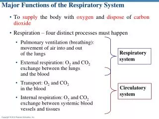
Exchange of Gases in the Lungs • Blood flow route • Right atriumright ventriclepulmonary arterylungsgas exchangepulmonary veinleft atriumleft ventricleaorta • Because of the thinness of the capillary wall, gases can be exchanged • Oxygen into the bloodstream to be distributed throughout the body • Carbon dioxide into the alveoli and then into exhaled air Respiratory System BIO 006
In the lungs, oxygen moves into the capillaries and combines with hemoglobin. It is transported in the bloodstream as oxyhemoglobin • Oxyhemoglobin breaks down into oxygen and hemoglobin and the oxygen moves out of the arterial capillary blood into tissue cells • Carbon dioxide moves from tissue cells into venous capillary bloodstream • Hemoglobin combines with carbon dioxide, forming carbaminohemoglobin. This will also travel to the lungs to be exhaled. Respiratory System BIO 006
Amount (volume) of Air Exchanges in Respiration • A spirometer can measure the volume of air exchanged in breathing • Tidal volume--amount normally breathed in or out with each breath • Inspiratory reserve volume—amount of air that can be forcibly inhaled after a normal inspiration • Expiratory reserve volume —amount of air that can be forcibly exhaled after expiring the tidal volume • Vital capacity —maximal volume you can exhale after a maximal inspiration • Residual volume—air that remains in the lungs after the most forceful expiration • Rate—usually about 12 to 18 breaths per minute; much faster during exercise Respiratory System BIO 006
Regulation of Respiration • Regulation of respiration permits the body to adjust to varying demands for oxygen supply and carbon dioxide removal • Respiratory control centers are located in the medulla and the pons. Centers in the pons have a modifying function. • Most important central regulatory centers are in the medulla; are called the inspiratorycenter and expiratory center • Under resting conditions, nervous activity in the respiratory control centers produces a normal rate and depth of respirations Respiratory System BIO 006
Respiratory control centers in the medulla are influenced by “inputs” from receptors located in other body areas: • Cerebral cortex—voluntary (but limited) control of respiratory activity • Chemoreceptors respond to changes in carbon dioxide, oxygen, and blood acid levels—located in carotid and aortic bodies • Pulmonary stretch receptors—respond to the stretch in lungs, thus protecting respiratory organs from overinflation Respiratory System BIO 006
Types of Breathing • Eupnea—normal breathing • Hyperventilation—rapid and deep respirations • Hypoventilation—slow and shallow respirations • Dyspnea—labored or difficult respirations • Apnea—stopped respirations • Respiratory arrest—failure to resume breathing after a period of apnea Respiratory System BIO 006
Disorders of Respiratory System Reduced air flow: • Asthma: causes partial closure of the bronchi and increased production of mucus. • Emphysema is caused by damage to the alveoli due to damage in the connective tissue in the bronchioles. The airways tend to collapse and this causes increased pressure in the lungs which eventually damage the alveoli. • Bronchitis is an inflammation of the bronchi which causes increased mucus which causes increased coughing. Respiratory System
Infections: • Pneumonia is an infection which causes inflammation of the lungs. The alveoli secrete excess fluid so gas exchange is impaired. • Tuberculosis is a bacterial infection which scars the lungs. • Botulism is a poisoning by bacterial toxin. The toxin blocks the transmission of nerve signals to the respiratory muscles. • Lung cancer • Congestive heart failure impairs lung functioning. • Cystic fibrosis is an inherited condition which causes mucus producing cells in the lungs to produce a very thick, sticky mucus which causes frequent infections. Other organs of the body are also involved. Respiratory System
Structure-Pharynx • Also called the throat • Divided into nasopharynx (portion behind the nose), oropharynx (portion behind the mouth), and laryngopharynx (portion behind the larynx) • Both nasal cavities, the mouth, the esophagus, the larynx, and the auditory (Eustachian) tubes open into the pharynx • Pharyngeal tonsils and openings of auditory tubes are into pharynx Respiratory System BIO 006
Diffusion is the process which is involved in gas exchange (passive movement from an area of high concentration to low concentration) Respiratory System BIO 006