Download
1 / 21
220 likes | 344 Vues
The Respiratory System rev 1-13. The primary function of the respiratory system is to deliver oxygen (O 2 ) to and remove carbon dioxide (CO 2 ) from the blood. The respiratory system also plays a role in maintaining the blood pH (acid-base balance).

E N D
The Respiratory System rev 1-13 • The primary function of the respiratory system is to deliver oxygen (O2) to and remove carbon dioxide (CO2) from the blood. • The respiratory system also plays a role in maintaining the blood pH (acid-base balance). • Additionally, in humans and most animals, the respiratory system also produces sounds. BIO 102 Respiratory System HANDOUT
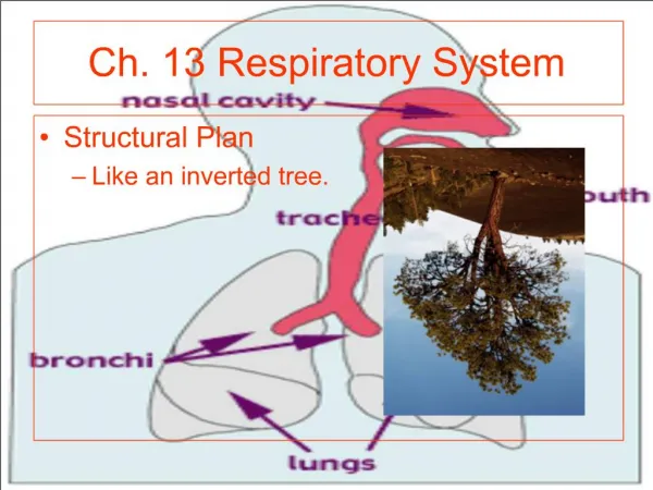
Anatomy of the Upper Respiratory Tract: • Nose, nasal cavities, sinusesand pharynx (throat) (everything above the Adam’s apple) • nose, nasal cavities and sinuses provide a large area of highly vascularized tissues which warm, filter and add moisture to air. • As air comes into contact with the warm, moist tissue of the nasal passages, it is warmed and moistened. The sinuses also add moisture to the air. • nose contains receptors for smell BIO 102 Respiratory System HANDOUT
The pharynx (throat) connects the nasal cavity and mouth to the larynx (voice box). • The tract provides a resonating chamber that gives your voice its characteristic tone Other structures which enter or are located in the pharynx are: • 2 tear ducts which carry fluid away from the eyes (this is why excess tears also make your nose runny) • the esophagus--this makes it possible to breathe through your mouth. BIO 102 Respiratory System HANDOUT
The 2 Eustachian tubes that drain the middle ear and equalize air pressure between the middle ear and outside air. • Food • Below the throat, the air passage crosses in front of the esophagus. This makes it possible for food or liquids to be accidentally sucked into the air passages and can cause us to cough or choke. These actions attempt to clear the food or liquid. BIO 102 Respiratory System HANDOUT
Epiglottis-a flap of cartilage located in the back of the throat. • During swallowing, the epiglottis forms a tight seal over the trachea so food can’t go down it. • The Uvula-a flap of tissue in the back of the mouth that hangs from the roof of your mouth. • This closes the upper air passages so food does not come out your nose. (This is also the part of the body that causes snoring when air passes over it.) BIO 102 Respiratory System HANDOUT
The lower respiratory tract includes • the larynx, trachea, 2 bronchi, 2 lungs (including the bronchioles and alveoli) • the larynx or voice box (nicknamed the Adam’s apple) is below the epiglottis and pharynx and is protected by the thyroid cartilage. • Functions of the larynx • maintains an open airway • route food and air into their appropriate tubes • assist in the production of sound BIO 102 Respiratory System HANDOUT
The vocal cords consist of 2 folds of connective tissue that extend across the airway. The opening of this airway is called the glottis. • Vocal cords are supported by ligaments. Sound is produced as we expel air past the stretched cords causing them to vibrate. BIO 102 Respiratory System HANDOUT
The trachea (or windpipe) is a tube below the larynx and transports air on its way into the lungs. • It is about 4 1/2 inches long, • is composed of C-shaped rings of cartilage (to ensure that it stays open), and carries air to the bronchi and through them to the lungs • The trachea branches into airways which are called the right and left bronchi (sometimes called the primary bronchi). These further subdivide into smaller and smaller bronchi. BIO 102 Respiratory System HANDOUT
The walls of the bronchi contain fibrous connective tissue and smooth muscle reinforced with cartilage. As the branches get smaller, the amount of cartilage declines. When they have no cartilage, their name changes into bronchioles. • Surrounding the bronchi are the lungs. These fill the thoracic cavity and extend from the clavicles to the diaphragm(a thin sheet of muscle). • Bronchioles lead to alveoli which are the air sacs of the lungs. Alveoli are composed of a single layer of flat, simple squamous cells and this is where gas exchange takes place. BIO 102 Respiratory System HANDOUT
Breathing • Involves repetitive cycles of getting air into and out of the lungs. • This requires muscular effort. • Since the lungs themselves do not have any skeletal muscle tissue, expansion and contraction occurs because the surrounding bones and muscles expand the size of the chest cavity. BIO 102 Respiratory System HANDOUT
Inspiration or Inhalation: • As the diaphragm contracts and flattens, the external intercostal muscles contract and lift the ribcage. This causes a pressure drop in the thoracic cavity. • The scalene and sternocleidomastoid (SCM) muscles also contract to help expand the thoracic cavity space. • As the volume (space) in the thoracic cavity increases, air rushes in to fill this space. BIO 102 Respiratory System HANDOUT
Other things that help inspiration: • The lungs and chest cavity are surrounded by a membrane called “pleura”. There is fluid between the layers of the pleura (pleural cavity) so the lungs can stretch and contract with minimum friction. • There is also a partial vacuum between the 2 pleural layers. This causes the lungs to stick to the chest wall as it expands. • Alveolar surfactant, a chemical within the lungs, decreases the surface tension so the lung tissue doesn’t stick to itself. BIO 102 Respiratory System HANDOUT
Expiration or Exhalation: • The diaphragm relaxes and intra-abdominal pressure pushes the diaphragm up. The internalintercostalmusclesand gravity help to drop the ribcage and thoracic cavity back to its smaller size. This increases pressure within the lungs and forces the air out of them. BIO 102 Respiratory System HANDOUT
Respiratory Volumes • Tidal volume is the amount of air an individual normally inhales and exhales. • Our body's normal breathing strategy is to ventilate the air sacs and also keep a minimal residual volume in the lungs. This allows us to keep some air for the blood passing through the lungs between breaths. This air is referred to as dead space volume. BIO 102 Respiratory System HANDOUT
The amount of air that can be forcibly inhaled after a normal inspiration (tidal volume) is called inspiratory reserve volume. • The amount of air that can be forcibly exhaled after a normal expiration (tidal volume) is called expiratory reserve volume. • The vital capacity is the maximal volume that you can forcibly exhale after a maximal inhalation. • After you forcibly exhale, there is always some air left in the lungs. This is called the residual volume. BIO 102 Respiratory System HANDOUT
These lung capacities are measured with a spirometer. • Gases are transported from the lungs to the body primarily by hemoglobin. They can also be dissolved in the plasma. In plasma, carbon dioxide dissolves and becomes carbonic acid or bicarbonate. BIO 102 Respiratory System HANDOUT
Gas Exchange and Transport: A Passive Process Gases diffuse according to their partial pressures (pressure exerted by 1 particular gas in a mixture of gases). No ATP is used. • External respiration: gases exchanged between air and blood • partial pressure of O2 is higher in alveoli-- CO2 is higher in the capillaries, so gases passively diffuse; • O2 carried to the blood and then to the body, CO2 exhaled • Internal respiration: gases exchanged with tissue (interstitial) fluids/space • partial pressure of O2 is lower in blood and CO2 is higher in the extracellular space, so gases passively diffuse; • O2 goes into the cell, CO2 diffuses into capillaries BIO 102 Respiratory System HANDOUT
Gas Exchange and Transport: A Passive Process cont’d • Oxygen transport: either bound to hemoglobin in red blood cells (primary method by which oxygen is transported) or dissolved in blood plasma • Forms oxyhemoglobin; depends on partial pressure of O2 (when P O2 rises, oxygen attaches to hemoglobin; when P O2 falls, oxygen detaches), temperature (likes lower temperature) & pH (likes neutral pH) • Hemoglobin’s affinity for oxygen is decreased by carbon monoxide • Carbon dioxide transport: dissolved in RBC and blood plasma in the form of bicarbonate (primary method by which CO2 is transported) or bound to the amino groups on the polypeptide chains of hemoglobin molecules • Carbaminohemoglobin, CO2 + H2O= H2 CO3 (carbonic acid)dissociates into HCO3-(bicarbonate) + H+ BIO 102 Respiratory System HANDOUT
Regulation of Breathing: Nervous System Involvement The nervous system regulates the rate and depth of breathing in order to maintain homeostasis. • The respiratory center in the medulla oblongata establishes basic breathing pattern including the rate at which we breathe. ---rate and depth of breathing is determined by the need to get rid of CO2 Chemical receptors monitor carbon dioxide, hydrogen ions, and oxygen levels: • The medulla is sensitive to hydrogen ions in cerebrospinal fluid as a result of more carbon dioxide in the blood. • Carotid and aortic bodies in the blood are sensitive to carbon dioxide, pH, and oxygen levels • Conscious control of breathing resides in higher brain centers (primarily the cortex)--ability to modify breath BIO 102 Respiratory System HANDOUT
Disorders of Respiratory System Reduced air flow: • Asthma: chronic inflammatory disorder of airways characterized by inflammation, bronchial hyper-responsiveness and airflow obstruction • causes constriction of bronchi, edema and increased production of mucus with possible mucus plugs. • COPD-Chronic Obstructive Pulmonary Disease • Emphysema caused by damage to alveoli due to damage in connective tissue in bronchioles; airways collapse and cause increased pressure in lungs which eventually damage the alveoli. • Bronchitis is an inflammation of the bronchi which causes increased mucus which causes coughing; Can be acute or chronic BIO 102 Respiratory System HANDOUT
Infections: • Pneumonia is an infection which causes inflammation of the lungs. The alveoli secrete excess fluid so gas exchange is impaired. • Tuberculosis is a bacterial infection causing lung scars. • Botulism is a poisoning by bacterial toxin. The toxin blocks the transmission of nerve signals to the respiratory muscles. • Lung cancer • Congestive heart failure impairs lung functioning. • Cystic fibrosis is an inherited condition which causes mucus producing cells in the lungs to produce a very thick, sticky mucus which causes frequent infections. Other organs of the body are also involved. BIO 102 Respiratory System HANDOUT