Download

1 / 37
390 likes | 536 Vues
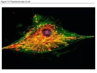
Figure 7.0 Fluorescent stain of cell. How do we learn about cell structure and function?. Immunofluorescence microscopy. http://learn.hamamatsu.com/galleries/digitalvideo/index.html. Figure 7.3 Cell fractionation. Differential Centrifugation - based on size (pellet and supernatant)

E N D
Figure 7.0 Fluorescent stain of cell How do we learn about cell structure and function? Immunofluorescence microscopy
http://learn.hamamatsu.com/galleries/digitalvideo/index.html
Figure 7.3 Cell fractionation Differential Centrifugation - based on size (pellet and supernatant) Density Gradient Centrifugation - Rate Zonal- preformed density gradients(size and shape) Equilibrium- Density
Figure 7.0 Fluorescent stain of cell How big is a cell?
What are the two main types of cells? Figure 7.4 A prokaryotic cell
Common Components of all Cells -molecular components -plasma membrane -DNA -cytoplasm -ribosomes -metabolism Animal Plant Bacteria (Prokaryotic) (Eukaryotic)
Figure 7.9 The nucleus and its envelope Nucleolus- site of ribosome synthesis Lamina- net of intermediate filaments Matrix- Structural fibers extending throughout nucleus
Figure 7.11 Endoplasmic reticulum (ER) Endomembrane System- internal membranes related by physical continuity or vesicle transfer. (nuclear envelope, E.R., golgi, lysosomes,and various vacoules) R.E.R.- synthesis and modification of excreted proteins, membrane proteins (glycoproteins). Vesicle transport to golgi. Membrane production. Smooth E.R.- Synthesis of lipids, carbo metabolism(glycogen hydrolysis), detoxification of poisons, Ca++ storage in muscles
Figure 7.10 Ribosomes Ribonucleoprotein complex- rRNA and protein
Figure 7.12 The Golgi apparatus Products from the E.R. modified, sorted, packaged for “shipping”. Polysaccharide synthesis (pectins in plants). “Docking proteins” in trans face membrane.
Figure 7.13 Lysosomes Sac of hydrolytic enzymes for all macromolecules. Bud from E.R. Acidic pH- H+ pumps in membrane.
Figure 7.14 The formation and functions of lysosomes (Layer 1)
Figure 7.14 The formation and functions of lysosomes (Layer 2)
Figure 7.14 The formation and functions of lysosomes (Layer 3) Digestion functions: -Food -Cell parts -Programmed cell death. Lysosome storage diseases: Tay-Sachs
Figure 7.16 Review: relationships among organelles of the endomembrane system
Figure 7.19 Peroxisomes Dehydrogenation reactions, formation of hydrogen peroxide. Peroxisomes not part of endomembrane system.
Figure 7.15 The plant cell vacuole Central vacuole: storage of macromolecules, inorganic ions, hydrostatic pressure. Contractile vacuole: Freshwater protists Pigment storage: Plastids
Figure 7.20 The cytoskeleton Structural support, cell motility, organelle movement and anchoring, intra-cellular transport, phagocytosis, regulation of biochemical activities (signal transduction). Not permanent, can disassemble and reassemble.
Figure 7.23 A comparison of the beating of flagella and cilia
Figure 7.24 Ultrastructure of a eukaryotic flagellum or cilium Basal body (Structurally like centriole)
Figure 7.26 A structural role of microfilaments Increase surface area Outer cytoplasmic area has gel consistancy.
Figure 7.27 Microfilaments and motility Distribution of nutrients and materials.
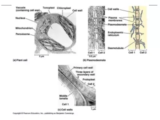
CELL SURFACES AND JUNCTIONS Matrix of microfibrils(cellulose), other polysaccharides and protein. Pectins (middle lamella)
Figure 7.29 Extracellular matrix (ECM) of an animal cell fibronectin
Figure 7.31 The emergence of cellular functions from the cooperation of many organelles