Download
1 / 23
230 likes | 239 Vues
Quantification of fluorescent signals Sabine Mai, Ph.D. Manitoba Institute of Cell Biology. Goals: Reliable analysis of data Comparison of fluorescent and molecular data Valid conclusions Precision of data Special goals for this workshop:

E N D
Quantification of fluorescent signals Sabine Mai, Ph.D. Manitoba Institute of Cell Biology
Goals: Reliable analysis of data Comparison of fluorescent and molecular data Valid conclusions Precision of data Special goals for this workshop: Rules of quantifications - relationship to other methods - relate this experience to own research program..
Reliable analysis of data using fluorescence Qualitative vs.quantitative analysis FISH signals: how many and where? how intense (deletion vs. amplification) Protein signals: where? How many? How much protein is expressed? Controls: what are appropriate controls?
Comparison of fluorescent and molecular data FISH: how does it relate to other techniques? How can it be compared? Single cell vs. whole cell population. Precision and combination with other techniques. Morphology. Protein: how does it relate to other techniques? Protein analysis of individual cells and of populations Protein localization studies. Precision. Morphology.
Valid conclusions. Data can be measured and compared between research and clinical laboratories. Data are stored and archived and can be revisited any time. Specific software makes analysis independent of user: valid, reliable, objective.
Precision of data. • Individual genes, • protein localization and movement, • chromosome structure and changes, • three-dimensional (3D) localization within the • interphase nucleus.
Spectral Imaging Qualitative analysis: genomic rearrangements.
Controls: SKY on primary cells (normal cells). Additional methods: M-FISH, painting, FISH, Southern.
Three-dimensional analysis of the interphase nucleus. Trisomy 11. Measurement of relative positions within the nucleus in mm scale.
Controls: Pirmary cell with normal chromosome 11. Additional methods: None.
Chromosome painting. Measurement of length of duplicated chromosome bands.
Controls: Normal cells (primary cells) without chromosomal changes. Additional methods: Southern blot
Centromere FISH. Evaluation of centromere numbers and sizes.
Controls: Internal control for centromere length Additional methods: Cytometry analysis of centromere length by FACS.
Q-FISH. Measurement of telomere length.
Controls: Internal control for telomere length Additional methods: Southern blot to measure relative telomere length Cytometry analysis of telomere length by FACS.
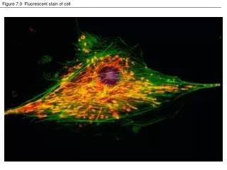
Fluorescent immunohistochemistry. Measurement of protein levels and protein location.
Controls: - Antibody controls - Cells that serve as positive and negative controls. Additional methods: Western analysis.
Fluorescent immunohistochemistry. Single cell and population analyses.
Controls: - Antibody controls - Cells that serve as positive and negative controls. Additional methods: Western analysis. Disadvantages: - no single cell analysis, only population analysis; - simultaneous analysis of several parameters is not possible; - localization of protein(s) is not as obvious.Contamination issues; - morphology of cell(s) is absent in Western blot.
Goals for the workshop: • Acquisition of images and analysis on different workstations. • Focus today: telomeres, protein, and later 3D analysis. • Analysis/measurements with different software packages: • Northern Eclipse • Applied Imaging • Teloquant
Please see Kim for the distribution of groups at different workstations and computers.