Download
1 / 50
520 likes | 964 Vues
Uveitis and Systemic Disease. Classification When to investigate ? Common Causes Systemic associations. Uveitis and Systemic Disease. Uveitis.

E N D
Uveitis and Systemic Disease Classification When to investigate ? Common Causes Systemic associations
Uveitis • Uveitis, a term correctly used to describe inflammation of the uveal tract (iris, ciliary body, choroid) alone, in reality comprises a large group of diverse diseases affecting not only the uvea but also the retina, optic nerve and vitreous. Uveitis is a major cause of severe visual impairment and has been estimated to account for 10-15% of all cases of total blindness in the USA. In surveys of the causes of blindness uveitis has usually not been included and is probably underestimated
Complications from chronic uveitis Complications from chronic uveitis are common and may result in severe visual loss.. Macular oedema can complicate any type of uveitis and can cause substantial visualloss. • Cataract is common in chronic uveitis and its treatment with corticosteroids. Techniques for cataract surgery and perioperativemanagement have improved greatly, and most patients with uveitisare now suitable for intraocular lens implantation and do well.18 • Glaucoma is the most overlooked complication of chronic uveitis and has several causes.19 Medical management with topicalagents such as blockers control the elevation of intraocularpressure in most patients. Some patients also require oral carbonicanhydrase inhibitors, while surgical intervention is reservedfor those who have progressive visual loss or uncontrollable intraocular
When to investigate • One of the most pressing questions that arises in the mind of every ophthalmologist who sees a new case of uveitis is "what is the cause of this disease?" In evaluating patients with uveitis, the ophthalmologist must consider that a lengthy list of infections, autoimmune systemic diseases, distinctive inflammatory conditions and masquerade syndromes may all cause uveal inflammation. Despite this array of potential diagnoses, the vast majority of patients have disease that defies categorisation.
Uveitis and Systemic Disease-avoid a shotgun approach to investigation !!Do not wade in like John Wayne !!
General Investigations • A recent retrospective review of patients with various types of uveitis showed the following abnormal results: full blood count: 23/113 (20.3%), plasma viscosity / ESR: 37/108 (34.2%), VDRL/TPHA: 3/70 (4.3%), angiotensin converting enzyme (ACE): 9/77 (10.8%) and chest x-ray (CXR): 15/103 (14.6%). Sarcoidosis was diagnosed in eight patients who had an abnormal CXR ± raised ACE. • All patients with symptoms of other organ system dysfunction or general malaise should be investigated to rule out under-lying systemic disease.
HLA-B27 disease. • Debate exists as to whether patients with the commonest type of uveitis (acute anterior uveitis - AAU) should be investigated. It is well recognised that approximately 50% of patients with AAU are HLA-B27 positive. A number of these patients will give a history of an associated HLA-B27 disease. • HLA-B27-associated AAU often presents with a number of clinical clues which help in diagnosis: it is usually recurrent, unilateral but alternating, with severe anterior chamber inflammation (posterior synechiae, fibrin and hypopyon).
Useful investigations for chronic uveitis • Chest x ray Diagnosis of tuberculosis, sarcoidosis, lymphoma, lung carcinoma • Syphilis serologyDiagnosis of syphilis • HLA-A29Diagnosis of birdshot chorioretinopathy • Mantoux testAnergic response despite prior BCG vaccination is consistent with sarcoidosis. Strong positive response without prior vaccination suggests exposure to tuberculosis • HIV serologyIf patient of high risk status or clinical picture suggests HIV related uveitis such as cytomegalovirus retinitis • Lyme disease serologyIf patient from endemic area or with history of exposure and suggestive symptoms • Antinuclear antibodiesIf clinical picture suggests juvenile chronic arthritis ANF ANCA Rhem Factor • Aqueous and vitreous biopsiesDiagnosis of infective endophthalmitis and intraocular lymphoma
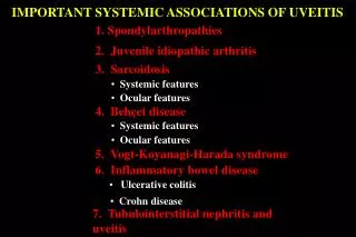
Uveitis and Systemic Disease Ankylosing Spondylitis 30% of AS patients develop iritis, especially if male; iritis may precede arthritis rarely retinal vasculitis / vitritis. Acute anterior uveitis lasting 2-6 weeks, good prognosis Investigations in suspected ankylosing spondylitis X-ray sacroiliac joints HLA B27 (positive in more than 90% )
Uveitis and Systemic Disease • Associations of Reiter's Syndrome • Occurs if genetically predisposed (HLA B27); 60 - 90% association • M>F • Exposure to certain urethritis / dysentery organisms: e.g. • Chlamydia, Yersinia, Shigella, Salmonella, Campylobacter. • The order of manifestation is normally: Œ urethritis conjunctivitis arthritis. • Ocular • 20% anterior uveitis, • 60% conjunctivitis, • episcleritis, keratitis, post-uveitis. • Reiter’s disease can sometimes result in hypopyon formation
Uveitis and Systemic Disease Sarcoidosis This chronic non-caseating granulomatous systemic disease of unknown aetiology affects women more commonly than men and is more common in individuals of Afro-Caribbean ethnicity. In Britain sarcoidosis is the commonest systemic disease that presents as chronic uveitis. It has protean ocular manifestationsand may present with a spectrum of ocular signs, including anteriorand posterior uveitis, retinal vascular sheathing, and optic discabnormalities Ocular Manifestations About 30% of patients with sarcoidosis have ocular involvement. Iritis may be acute or chronic; it may be unilateral or bilateral. Patients with posterior uveitis usually have anterior uveitis as well. Vitritis is also common and tends to occur in older patients. There may be retinal periphlebitis; the vessels may display an exudative cuff (so called ‘candle wax drippings’). Inflammation of the retina may lead to macular oedema, retinal granuloma, preretinal nodules and retinal haemorrhage. Inflammation of the optic nerve may cause optic disc oedema, granuloma and neovascularization. Branch retinal vein occlusion and retinal neovascularisation are uncommon
Uveitis and Systemic Disease Sarciodosis - Investigations Chest X-ray Serum ACE (angiotensin converting enzyme)- this is elevated in active disease urine and serum calcium levels- hypercalciuria is common hypercalcaemia is less common Conjunctival biopsy may show granulomata
Uveitis and Systemic Disease Ocular Manifestations of Tuberculosis Affects 2% of sufferers of active tuberculosis , uveitis is commonest manifestation. Systemic disease is often apparent. Eyelids- lupus vulgaris (nodules surrounded by erythema) Orbit- cellulitis, dacryoadenitis, dacryocystitis, osteomyelitis, abscess Conjunctiva- rarely affected, chronic conjunctivitis Cornea- phlyctenular keratoconjunctivitis, interstitial keratitis (unilateral, sectorial, superficial vascularisation) Sclera- episcleritis, nodular scleritis Uveitis- chronic granulomatous anterior uveitis, multifocal choroiditis, exudative retinitis, vasculitis, optic nerve oedema, papilloedema
Uveitis and Systemic Disease Juvenile Chronic Arthritis Chronic AAU , usually bilateral Commoner in female patients, the young, ANF positive. Pauciarticular disease <5 joints. Complications Glaucoma (20%) Cataract (40%) Band Keratopathy (40%)
Uveitis and Systemic Disease Monitoring Children with Juvenile Chronic Arthritis High Risk Early Onset , < 6 years, Pauciarticular Disease , ANA Positive 3 months for first year , then 6 months for five years , then annually Medium Risk polyarticular disease ANA positive , pauciarticular -disease ANA negative 6 monthly intervals for 5 years then annually Low Risk Systemic JCA , B27 associated arthritis , disease starting after age 11 Duration For ten years after onset of JCA or until age 12, whichever is shorter. Source RCOphth (UK), British Paediatric Association (1994)
Masquerade Syndromes Intraocular lymphoma may present as a chronic uveitis in older patients, especially when there is vitritis and vitreous veilsand a poor response to treatment. Intraocular tumours, particularlyretinoblastoma in children, may also occasionally present in thismanner. Differential Diagnosis Of Uveitis- It is of paramount importance to note that uveitis can be caused or mimicked by the following- “Masquerade Syndromes”- neoplasms mimicking uveitis Ocular malignant melanoma Retinoblastoma Reticulum Cell Sarcoma ( Primary Intraocular Lymphoma ) Leukaemia Lymphoma Ocular Metastasis Other- Endophthalmitis Retinal detachment Intraocular foreign body
Uveitis and Systemic Disease Syphilis Uveitis may be acute or chronic, unilateral or bilateral. Interstitial keratitis affects a small percentage of acquired cases and is often unilateral. Chorioretinitis is bilateral in 50% of cases;multifocal or diffuse yellow exudate is seen. The chorioretinitis may resolve, leaving extensive bone spicule pigmentation. The appearance may resemble retinitis pigmentosa. There may be retinal oedema, haemorrhages, exudates, cotton wool spots and vascular sheathing. Optic disc oedema may also be seen. Investigations for suspected syphilitic uveitis include VDRL and FTA-ABS tests. The VDRL test is useful for screening; false positive results may occur. The FTA-ABS test remains positive for life, even after treatment.
Uveitis and Systemic Disease- about 5% of uveitis caused by syphilis in some series
Therapeutics • The aims of treatment are to control inflammation, prevent visual loss, and minimise long term complications of the diseaseand its treatment. Macular oedema is the commonest indicationfor treatment. Treatment is usually indicated if the visual acuityhas fallen to less than 6/12, or if the patient is experiencingvisual difficulties. In patients with longstanding macular oedemaand poor vision or where it is not possible to determine easilythe cause of visual loss, a trial of immunosuppressive treatmentis usually indicated to determine whether the visual loss is reversible.Many patients with unilateral chronic uveitis can be managed withtopical corticosteroids to control anterior uveitis and periocularcorticosteroids for macular oedema and visual loss. Patients withuseful vision in only one eye must be managed aggressively tocontrol inflammation and preservevision.
Systemic corticosteroids • Corticosteroids are the mainstay of systemictreatment for patients with chronic uveitis, and the usual indicationfor treatment is the presence of macular oedema and visual acuityof less than 6/12. • Patients should be treated with appropriatedoses to determine whether the macular oedema is reversible. Thusmaximum treatment (1.0-1.5 mg/kg body weight/day of prednisoneor prednisolone) should be used for two to three weeks. • If thereis no response at this dose, addition of a second line agent suchas cyclosporin (or azathioprine or mycophenolate in older patients)for a further four to six weeks may be considered. In childrenthe doses should be adjustedappropriately.
Other systemic immunosuppressive therapy • If macular oedema recurs and visual acuitydecreases at an unacceptably high dose of corticosteroid (>15mg/day of prednisolone) an additional drug is necessary to helpcontrol the inflammation. Cyclosporin is the drug of choice formost patients aged under 50 years.The commonest doselimiting side effects of cyclosporin are hypertension and renaldysfunction, which are usually reversible if the drug isstopped. • Several other drugs can be considered in patients who require additional immunosuppressive therapy when cyclosporin is notappropriate or not tolerated. Azathioprine, methotrexate, and,much less commonly, cyclophosphamide are the most used, but eachis associated with important side effects and complications.Other agents such as mycophenolate, tacrolimus, and humanisedTac monoclonal antibodies have been usedThe decisionto start treatment with immunosuppressive drugs is a long termcommitment by both the clinician and patient, as treatment islikely to last for a minimum of six months and is often muchlonger.
Surgery • Surgery may be required for complicationssuch as cataract, glaucoma, and vitreoretinal problems, but, exceptin emergency situations, it should be contemplated only once theuveitis is controlled, ideally for at least three months. Intraocularsurgery (cataract removal, vitrectomy, and retinal detachmentsurgery) is performed under the cover of systemic corticosteroidsto prevent a relapse of uveitis. Removal of the vitreous body(vitrectomy) may be helpful when there is substantial opacitybut also may improve disease control, particularly in youngerpatients.
Complications of chronic uveitis and their management • Macular oedema • Periocular steroids • Systemic steroids • Immunosuppressive drugs • Cataract • Surgery once uveitis controlled for 3 months preoperatively • Perioperative cover with corticosteroid • Intraocular lens in most patients • Glaucoma • Management depends on typeTopical drugsShort term treatment with systemic carbonic anhydrase inhibitorsSurgery • Synechiae • Minimise with regular mydriatics • Band keratopathy • Chelation with EDTA • Excimer laser • Vitreous opacities • Observation • Occasionally short course of corticosteroids • Vitrectomy rarely required • Vitreous haemorrhage • Observation • Exclude new vessels and retinal tear as cause • Retinal neovascularisation • Control uveitis • Laser photocoagulation if ischaemia present • Subretinal neovascularisation • Observation • Laser photocoagulation • Interferon a • Surgical membranectomy • Retinal detachment • Determine whether exudative, rhegmatogenous, or traction • Surgery usually involves vitrectomy • Perioperative cover with corticosteroid
Summary points • Intraocular inflammation has various causes and can be acute or chronic • In either case the inflammatory process can be apparently localised to the eye or be part of a systemic disease such as sarcoidosis or Behçet's disease • The inflammation can occur in any part of the eyeanterior, posterior, or bothand visual loss can occur with any type • Treatment depends on the location and severity of the inflammation, with systemic drugs being reserved for sight threatening posterior disease • Complications are common and include cataract, glaucoma, macular oedemaall of which can reduce vision