Download
1 / 58
1.71k likes | 5.07k Vues
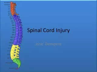
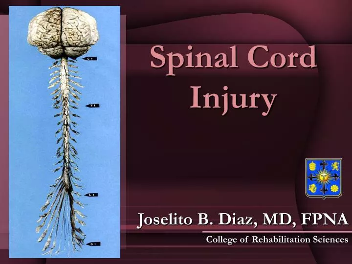
Spinal Cord Injury. Joselito B. Diaz, MD, FPNA College of Rehabilitation Sciences. Spinal cord. Continuation of the medulla Commences at the foramen magnum and extends to the level between L1 and L2 vertebrae 31 segments 8 cervical 12 thoracic 5 lumbar 5 sacral 1 coccygeal.

E N D
Spinal Cord Injury Joselito B. Diaz, MD, FPNA College of Rehabilitation Sciences
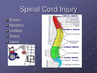
Spinal cord • Continuation of the medulla • Commences at the foramen magnum and extends to the level between L1 and L2 vertebrae • 31 segments • 8 cervical • 12 thoracic • 5 lumbar • 5 sacral • 1 coccygeal
Spinal cord • Posterior roots convey sensory inputs while the anterior roots conduct efferent axons to the innervate skeletal muscles • Arterial blood supply comes from the anterior spinal and 2 posterior spinal arteries • Anterior spinal artery supplies all the cord except the dorsal horn and posterior column
Spinal cord (descending tracts) • Lateral and anterior corticospinal tract – convey axons from the motor cortex • Vestibulospinal tract – convey axons from the lateral vestibular nucleus; facilitate activity of extensor muscles • Reticulospinal tract – transmit autonomic axons and respiratory motor fibers; facilitate or inhibit voluntary movement • Rubrospinal tract – facilitate activity of flexor muscles • Tectospinal tract – arise in the superior colliculus, serves as efferent pathway for spinal-visual reflexes
Motor pathway Spinal effector Mechanism
Spinal cord (ascending tracts) • Lateral spinothalamic tract – carry pain and temperature sensation • Anterior spinothalamic tract – pathways for crude touch and pressure • Posterior columns – consists of two tracts which convey position, discriminative touch and vibration sense • Fasciculus gracilis and cuneatus • Spinocerebellar tract – convey propioceptive sensation to the cerebellum
Dorsal column L T S C Corticospinal tract S Spinothalamic tract L T C C T L S
SCI Classification • Tetraplegia – results from injury in the cervical spinal cord • Paraplegia – results from injury in the thoracic, lumbar or sacral segments • The neurological level is the most caudal spinal segment that retains normal motor and sensory function on both sides of the body • The rectal area is examined digitally • A complete lesion is defined as the absence of sensory or motor function in the lower sacral segment • Incomplete lesion if either sensory or motor function is present and known as sacral sparing
ASIA defines the Zone of Partial Preservation as up to 3 segments caudal to the level of a complete injury, in which impaired sensory or motor function is found • If any function is found more than 3 segments below the level, the lesion is incomplete
Mechanisms of Injury 1. TRAUMA TO THE SPINE AND SPINAL CORD • 10-14% of spinal fractures and dislocations result in SCI • Fracture-dislocation, pure fracture and pure dislocation (3:1:1) • Direct blow to the spine is a relatively uncommon cause of serious spinal cord injury except for bullets, sharpnel and stab wounds • Mechanisms: • Vertical compression of the spinal column to which anteroflexion is added • The adjacent cervical vertebrae are forced together at the level of maximum stress and the anteroinferior edge of the upper vertebral body is driven into the one below, sometimes splitting it in two. The posterior part of the fractured body is displaced backward and compresses on the cord
Mechanisms of Injury • Vertical compression and retroflexion (hyperextension) • Stress is mainly on the posterior elements, which may fractured, and on the anterior ligaments. This allows for displacement of one vertebral body on the adjacent one. • Whiplash or recoil injury • Sudden forward flexion of the neck followed by retroflexion • Transient posterior dislocation of a vertebral body or retropulsion of the intervertebral disc into the spinal canal • High velocity missile penetrates the vertebral canal and damages the spinal cord directly
Mechanisms of Injury • Most common sites of vertebral injuries • 1st and 2nd cervical vertebrae • 4th to 6th cervical vertebrae • T11 to L2 vertebrae • Most mobile portions and the regions in which the cervical and lumbar enlargements greatly reduce the space between neural and bony structures
Mechanisms of Injury 2. Radiation injury of the spinal cord • Well recognized sequela of radiation therapy • Transient radiation myelopathy • Delayed progressive radiation myelopathy 3. Spinal cord injury due to electric currents and lightning 4. Myelopathy following lumbar puncture
Mechanisms of Injury 5. Spinal tumors 6. Demyelinating diseases (MS, transverse myelitis) 7. Infectious myelitis including Pott’s disease 8. Degenerative joint diseases e.g. cervical spondylosis 9. Vascular diseases of the spinal cord • Infarction • Hemorrhage of the spinal cord and spinal canal • Vascular malformations 10. Hereditary disorders e.g. familial spastic paraplegia 11. Congenital disorderse.g. tethered cord
Clinical syndromes Complete cord transection syndrome • Caused by fracture dislocation of the vertebral column, bullet or stab wound, or an expanding tumor • Bilateral flaccid paralysis and muscular atrophy at the level of the lesion • Bilateral spastic paralysis below the level of the lesion • Bilateral loss of all sensation below the level of the lesion (pain and temperature loss 1-2 segments below the lesion) • Loss of voluntary bowel and bladder control
Clinical syndromes Central cord syndrome • Generally occur in the cervical area • Occurs in elderly with DJD in the neck following hyperextension injury; syringomyelia • Greater UE spastic weakness than LE • Segmental loss of pain and temperature • Bilateral atrophic, areflexic paralysis due to involvement of the AHC • Bilateral loss of all sensations below the level of the lesion with characteristic “sacral sparing”
Clinical syndromes Brown-Sequard syndrome • Lateral hemisection of the cord e.g. bullet, knife wound, expanding tumor • Ipsilateral spastic paralysis below the injury • Ipsilateral loss of vibration and position sense • Contralateral loss of pain and temperature 1 or 2 segments below the injury • Ipsilateral flaccid paralysis at level of injury (AHC) • Ipsilateral paresthesia/radiculopathy/ anesthesia in the segment of the lesion (dorsal root) • Simple touch maybe unimpaired
Clinical syndromes Anterior cord syndrome • Thrombosis of the anterior spinal artery, herniated intervertebral disc, cord contusion during vertebral fracture or dislocation • Bilateral flaccid paralysis at the level of the lesion • Bilateral spastic paralysis and loss of pain and temperature sense below the lesion • Intact position and vibration sense
Clinical syndromes Posterior cord syndrome • Thrombosis of the posterior spinal artery or vertebral artery dissection (uncommon) • Surgery or trauma to the spine • Bilateral loss of position and vibration sense below the lesion • Motor function and sense of pain and temperature preserved
Stages in Complete Transection • Spinal shock • Lasts 1-6 weeks, average of 3 weeks • Due to loss of excitatory descending influences, the findings below the lesions are: • Loss of somatic sensation • Loss of visceral sensation • Loss of motor function • Loss of muscle tone • Loss of reflex activity
Stages in Complete Transection • Stage of Minimal Reflex Activity • 3-6 weeks • Characterized by: • Bulbocavernous reflex (heralds its onset) • Weak flexor responses to nociceptive stimuli which begin distally and later proximal
Stages in Complete Transection • Stage of Flexor Muscle Spasm (Spasticity) • 6-16 weeks • Characterized by: • Increasing tone in flexor muscles • Stronger flexor response to nociceptive stimuli which progressively involve more proximal muscle group • Triple flexion response – flexion at hip, knee and ankle with mild nociceptive stimulus or • Mass reflex response – mild stimulus leads to powerful bilateral triple response (due to spread of afferent impulses from one segment to the next and continue to fire even after the stimulus withdrawal)
Stages in Complete Transection • Stage of alternate flexor and extensor spasticity • More than 4 months • Both flexor and extensor spasms occur, but within relative short time, extensor muscle tone may be so great that patient can momentarily support his weight in standing position
Stages in Complete Transection • Stage of predominant extensor spasticity • More than 6 months • Characterized by: • Complete paralysis below lesion • Marked extensor spasticity • Loss of somatic/visceral sensation • Increased MSR • Bilateral Babinski • Clonus in both lower extremities • Bowel and bladder incontinence • Reflex spinal sweating in response to noxious stimuli
Complications of SCI • Neurogenic bladder • UMN type – lesion above S2 level • LMN type – lesion at S2-S4 • Cystometry offers the best diagnostic tool for differentiation UMN from LMN • Reflex tests to differentiate
Complications of SCI • Long term indwelling or suprapubic catheter complications • UTI (most common morbidity) • Epididymo-orchitis • Urethral diverticuli • Vesicovaginal or penoscrotal fistula • Bladder calculi • Intermittent catheterization program – retraining the bladder to function on its own
Complications of SCI • Methods of stimulating micturition • Stroking or tapping the upper medial thigh • Alternate hot and cold compress • Sound of water from the faucet • Gentle pulling of the pubic hair • Insertion of gloved finger into the anus to relax the sphincter • Residual urine determination • %RU = [RU/(RU+VU)] x 100 • If %RU is < 20%, trial with catheter out • If %RU is > 20%, reinsert indwelling catheter or continue ICP
Complications of SCI • Neurogenic bowel • Decreased intestinal activity • Diarrhea caused by fecal impaction • Bowel program • Food • High residue diet • Fruits • Fluid intake • 8-12 liters • Stool softeners • Timing • Postprandial (gastrocolic reflex) • Assists: suppositories, digital stimulation, laxatives • Regularity: early morning/evening • Positioning • Sitting down (physiologic position)
Complications of SCI • Autonomic hyperreflexia • Complication in lesions above T6 • Signs and symptoms • Pilomotor erection • Hypertension • Headache • Pallor • Dyspnea • Sweating • Management: • Since bladder distention is the most frequent cause, urinary drainage should be established immediately • Rule out causes from the intestinal tract • Cold extremities below the level of lesion • Bradycardia • Flushing above the level of lesion • Hot above the level of lesion
Complications of SCI • Spasticity • Aggravated by noxious stimuli below the level of the lesion e.g. skin lesions, ischemic muscle areas or irritation from the bowel and bladder • 3 characteristic patterns • Increased myotatic reflexes • Resistance to passive range of motion • Clonus and Babinski
Complications of SCI • To allow good circulation to insensitive muscles, self-stretching exercises are recommended • If spasticity interferes with functional activity, medications should be given • Diazepam, baclofen, tizanidine, dantrolene • Neurological procedures • Botulinum toxin injection • Intrathecal alcohol block, rhizotomy, myelotomy
Complications of SCI • Pressure ulcers (decubitus ulcers, bed sores, pressure sores) • Area of unrelieved pressure over a defined area, usually over a bony prominence, resulting in ischemia, cell death and tissue necrosis • Most important etiologic factor is localized ischemia due to excessive pressure • Secondary factors: sensory loss, paralysis, joint contractures, spasticity, incontinence
Complications of SCI • Sites of occurrence (depends on positioning of patient) • Ischial tuberosities (if in sitting position) • Greater trochanter (if in lateral decubitus position) • Sacrum (if lying on back) • Heels • Others: knees, lateral malleoli, scapulae, spinous process, occiput, head of fibula, ASIS and patella