Download
1 / 29
821 likes | 2.5k Vues
Nervous Regulation of Circulation and Rapid Control of Arterial Pressure. Autonomic Nervous System. Sympathetic Parasympathetic. Autonomic Nervous System. Sympathetic Innervation of Blood Vessels.

E N D
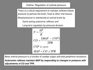
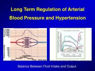
Nervous Regulation of Circulation and Rapid Control of Arterial Pressure
Autonomic Nervous System • Sympathetic • Parasympathetic
Sympathetic Innervation of Blood Vessels • Distribution of sympathetic nerve fibers to all blood vessels except the capillaries, precapillary sphincters and metarterioles • Innervation of large vessels, especially veins and increased venous return to the heart
Sympathetic Nerve Fibers to the Heart • Sympathetic stimulation markedly increases the activity of the heart • Both increases the heart rate and enhances its strength and volume of pumping.
Parasympathetic Control of the Heart Function • Parasympathetic plays a minor role in regulating of the circulation • Its most important circulatory effect is to control heart rate by way of parasymphatetic nerve fibers to the heart (N. Vagus)
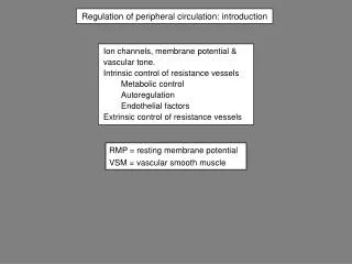
Sympathetic Vasoconstrictor System and Its Control by the CNS • The sympathetic nerves carry tremendous numbers of vasoconstrictor nerve fibers and only a few vasodilatatory fibers • This sympathetic vasconstrictor effect is especially powerful in the kidneys, intestines, spleen and skin but much less potent in skeletal muscle and the brain
Vasomotor Center in the Brain • Located in the reticular substance of the medulla and lower pons • Important areas in this center: • A vasoconstrictor area • A vasodilatator area • A sensory area, located in the nucleus tractus solitarius • Output signals from the sensory area provides a reflex control of many circulatory functions (e.g. baroreceptor reflex)
Sympathetic Vasoconstrictor Tone • Continuous partial constriction of the blood vessels is normally caused by sympathetic vasoconstrictor tone
Control of the vasomotor center by higher nervous centers • Large numbers of small neurons located throughout the reticular substance of the pons, mesencephalon and diencephalon can excite or inhibit the vasomotor center • Role of hypothalamus – Limbic system • Many parts of the cerebral cortex can also excite or inhibit the vasomotor center • vasovagal syncope (emotional fainting)
Sympathetic Vasoconstrictor Transmitter Substance • Noradrenalin • Alpha adrenergic receptors of the vascular smooth muscle • Adrenal medullae and their relation to the sympathetic vasoconstrictor system (adrenalin and noradrenalin) • Sympathetic vasodilator system and its control by the central nervous system • Skeletal muscles and vasodilator fibers…
Role of the nervous system in rapid control of arterial pressure • Rapid control of arterial pressure within 5-10 seconds • Stimulation of entire vasoconstrictor and cardioaccelerator functions by the sympathetic system • Almost all arterioles of the systemic circulation are constricted • The veins are strongly constricted • The heart itself is directly stimulated by the ANS, further enhancing cardiac pumping
Increase in AP During Muscle Exercise and Other Types of Stress • During heavy exercise, skeletal muscle require increased blood flow – role of local vasodilation • Increase of AP (30-40%) in heavy exercise increases blood flow • Activation of vasomotor center • Other types of stress and increased AP • Alarm reaction • Fight or flight
Maintaining Blood Pressure: Short Term Mechanisms - CNS • Baroreceptor initiated reflex • located at carotid sinuses and aortic arch • monitors blood pressure • regulates the activity of the sympathetic nervous system (vascular tone)
Baroreceptor Reflexes • This reflex is initiated by stretch receptors (baroreceptors or pressoreceptors) located at specific points in the walls of large systemic arteries • Carotic and Aortic baroreceptors • Signals from carotid baroreceptors – small Hering’s nerves – N. Glossopharyngeus – NTS in the medulla • Aortic baroreceptors – N. Vagus – NTS in the medulla
Response of Baroreceptors to Pressure • Carotid sinus baroreceptors are not stimulated by pressures between 0-50 mmHg, but above this they respond progressively more rapidly • Aortic baroreceptors are similar, but they operate 30 mmHg or higher pressures
Circulatory Reflex Initiated by the Baroreceptors • After the signals from baroreceptors enter the NTS, secondary signals inhibit the vasoconstrictor center and excite vagal parasympathetic center • The net effects are: • Vasodilation of the veins and arterioles in the peripheral circulatory system • Decreased heart rate and strength of the heart contraction • Increased TPR (total peripheral resistance) and decreased cardiac output
Circulatory Reflex Initiated by the Baroreceptors Ligation of two common carotid arteries
Baroreceptors and Changes in Body Posture • AP in the head and upper parts of the body tends to fall immediately on standing • This may cause loss of consciousness • Falling pressure at the baroreceptors elicits an immediate reflex resulting in strong sympathetic discharge • Pressure Buffer Function of the Baroreceptor control system • Reduction of minute by minute variations in arterial BP • Long term regulation of arterial BP
THE BARORECEPTOR REFLEX - AN EXAMPLE CORRECTION OF POSTURAL HYPOTENSION On standing up venous return falls Cardiac output diminishes Arterial blood pressure is reduced Baroreceptor afferent firing reduced Medullary centres inhibition reduced Effect of gravity on venous return Preload diminished - Starling’s Law Subject possibly feels faint as cerebral flow is reduced Due to reduced arterial B.P. Vasoconstriction Tachycardia Raised stroke work Tend to restore arterial blood pressure Increased sympathetic tone to arterioles Reduced vagal tone to s.a. node Increased myocardial sympathetic tone
Maintaining Blood Pressure: Short Term Mechanisms - CNS • Chemoreceptor initiated reflexes • Carotid bodies, aortic bodies • Monitor changes in indicator chemicals (O2, CO2, H+, HCO3-) • CO2, H+, O2 (stresses) result in sympathetic activity and BP
Control of the Arterial Pressure by the Carotid and Aortic Chemoreceptors • Abundant blood flow and contact with the chemoreceptors • Signals from the chemoreceptors excite the vasomotor center and this elevates the AP back to normal • Chemoreceptor reflex is not a powerful AP controller until the AP falls below 80 mmHg • In low pressures this reflex becomes important
Other Reflexes Regulating Blood Pressure • Atrial and pulmonary artery reflexes that help regulate AP and other circulatory factors: • Both atria and pulmonary arteries have in their walls stretch receptors called low-pressure receptors • Atrial reflexes that activate the kidneys (volume reflex) • Stretch of the atria also causes reflex dilation of afferent arterioles in the kidney • Atrial reflex control of the heart rate (Bainbridge reflex) • Increased atrial pressure also increases heart rate • Direct effect of increased atrial volume to stretch the sinus node • Additional 40-60% increase in rate is caused by a nervous reflex (Bainbridge reflex) that transmits afferent signals to the medulla of the brain
Central Nervous System Ischemic Response • Most nervous control of BP is achieved by baroreceptors, chemoreceptors and low-pressure receptors: These are all located in the peripheral circulaiton • However, cerebral ischemia causes strong excitation of the vasomotor center • Accumulation of carbon dioxide • Other factors (build up of lactic acid) • CNS ischemic response is one of the most powerful of all the activators of the sympathetic vasoconstrictor system • Importance of the CNS ischemic response • Activated only at 60 mmHg or below • Emergency pressure control system
Central Nervous System Ischemic Response A: Vasomotor waves caused by CNS ischemic response B: Vasomotor waves caused by baroreceptor reflex
Special Features of Nervous Control of Arterial Pressure • Abdominal compression reflex • Compression of large abdominal veins and other vessels by skeletal muscles of the body, especially abdominal muscles • Increased cardiac output and arterial pressure caused by skeletal muscle contraction during exercise • Compression of blood vessels by skeletal muscles
Respiratory Waves in the Arterial Pressure • 4 to 6 mmHg fall in AP during respiratory cycle • Breathing signals arise in the respiratory center of the medulla “spill over” into the vasomotor center with each respiratory cycle • With inspiration, pressure in thoracic cavity becomes negative allowing blood vessels in the chest to expand * This reduces the venous return and decreases the cardiac output • Pressure changes in the thoracic vessels by respiration can excite vascular and atrial stretch receptors